 English
 Pусский
 Français
 Español
 Portugues
 Deutsch
 lingua italiana
 العربية
 Melayu
 Filipino
 Монгол хэл
 dansk
dansk Bahasa indonesia
 日本語
日本語 한국어
한국어 Türk dili
 Tiếng Việt
 svenska
 Nederlands
 Norsk språk
|
What is Confocal Laser Scanning Microscope (CLSM)?
|
|
 |
 |
|
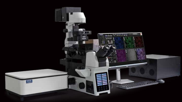
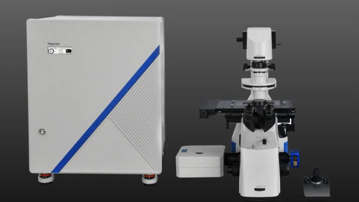
A64.0960
|
A64.1095
|
|
Key Word
Confocal
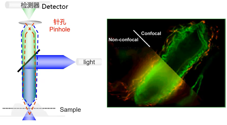
Confocal means "having the same focal point," The Light Source Pinhole and Detection Pinhole are conjugately focused to the objective focal plane.
Laser
Laser beams are used as a light source.
Scanning
Point-by-point imaging, line-by-line scanning, layer-by-layer scanning.
Theory
CLSM microscope creates an image by only detecting the light that originates from the same focal point as the laser beam, while excluding the light from other planes. The confocal design of the microscope provides several advantages over conventional microscopes, such as improved depth resolution, reduced background blur, and the ability to generate 3D reconstructions of the specimen.
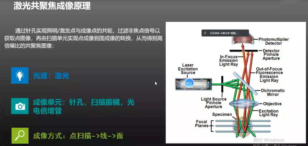 What is Confocal Laser Scanning Microscope (CLSM)? A Confocal Laser Scanning Microscope (CLSM) is an optical imaging technique uses lasers to generate images with high resolution and 3D reconstruction capabilities. It is consisted by a fluorescent microscope and laser scanning device. It works by illuminating a sample with a focused laser beam passing Light Source Pinhole, excite the sample to emit fluorescence, and then capturing the emitted fluorescence of the sample using a Detectio Pinhole aperture placed in front of the detector. This process helps to eliminate out-of-focus light to create a precise image with high contrast and resolution. CLSM is widely used in biology, medicine, materials science, and physics research.
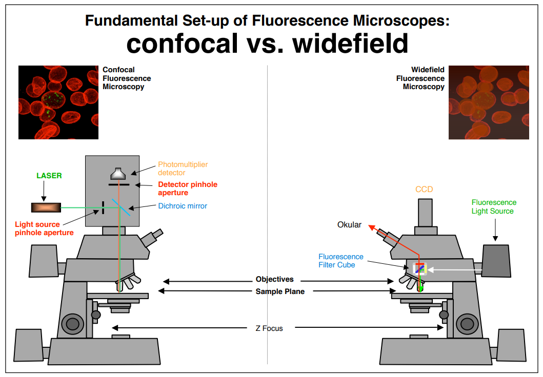 Traditional fluorescence microscopes use fluorescent substances to label specific structures in cells, which not only enhances the contrast between the image and the background, but also greatly improves the resolution (δ=0.61·λ/NA, where δ is the resolution of the microscope; λ is the wavelength of the illuminating light; and NA is the numerical aperture of the objective lens) since many fluorescence microscopes use short-wavelength ultraviolet light as the light source. However, a difficult-to-overcome drawback of traditional fluorescence microscopes becomes apparent when the observed fluorescence sample is slightly thicker: the fluorescence structures outside the focal plane become blurry and vague. This is because most biological samples are multi-layered, three-dimensional structures (such as the basilar membrane of the cochlea, which is a spatial structure composed of outer hair cells, various supporting cells, and nerve fibers). Focusing on different planes with a regular optical microscope will reveal different shapes. If the fluorescently labeled structures are distributed across different layers and overlap with each other, the epifluorescent microscope not only collects light from the focal plane, but also scatters the fluorescence from above or below the focal plane, which greatly reduces the optical resolution of the fluorescence microscope.
|
|
 |
 |
|
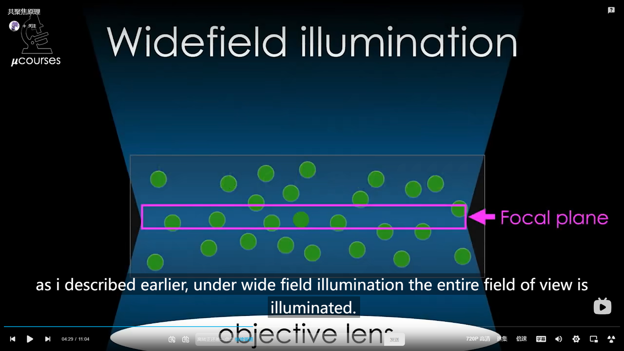
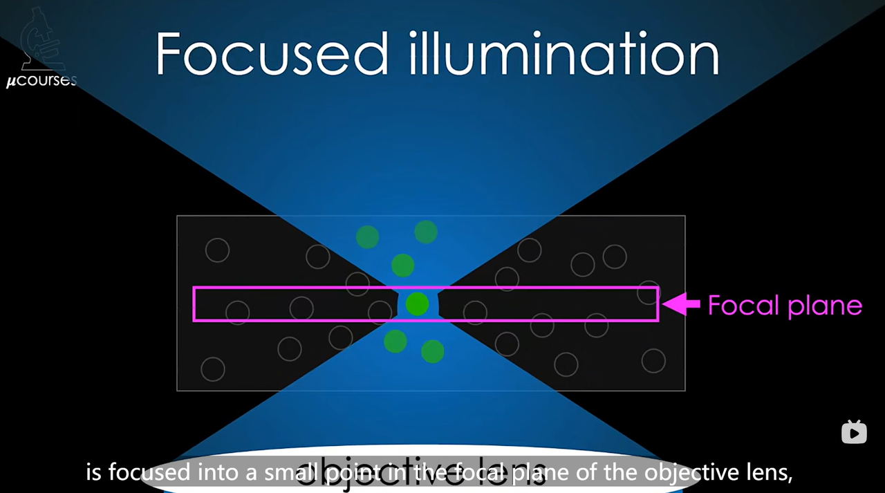
The function of pinhole: Wide Field vs. Focused
|
|
|
Based on traditional optical microscopes, laser scanning confocal microscopy uses laser as the light source, adopts the principle and device of conjugate focusing, and the points outside the focal plane do not image at the detection pinhole. It uses computer to perform digital image processing for observation, analysis and output of the observed objects. Its features include being able to perform section scanning and 3D imaging of the sample, and non-destructively observing and analyzing the three-dimensional spatial structure of cells.
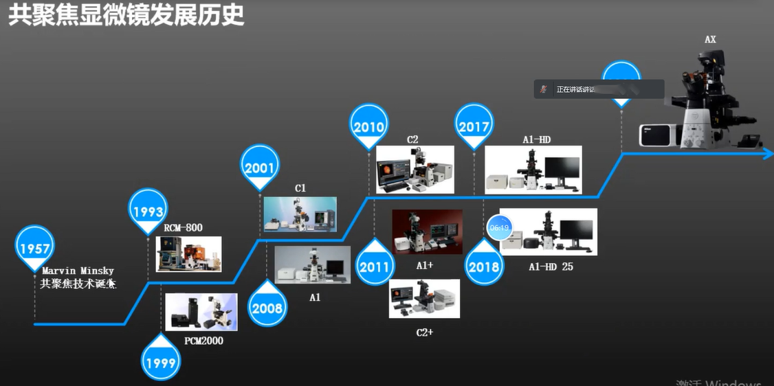
 Development History
The principle of confocal microscopy was first proposed by Marvin Minsky, an American cognitive scientist and computer scientist, in 1957. He envisioned using a beam of light focused on a single point to illuminate a sample, which could then be scanned point-by-point to generate an image.
This concept was later refined by a group of researchers led by Robert Campbell at the University of Rochester in the 1970s, who developed the first confocal microscope prototype. They used a spinning Nipkow disk to filter out out-of-focus light and capture images with greater resolution and clarity compared to traditional microscopy methods.
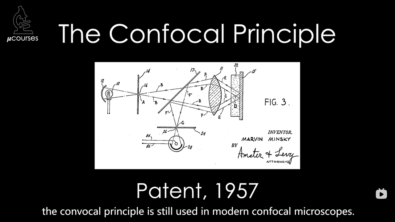 Since then, the development of confocal microscopy has continued, with new advancements in laser technology, computer processing, and image analysis software. Today, confocal microscopy is widely used in many different fields of research, from biology and medicine to materials science and engineering.
 Application
Confocal laser scanning microscopy (CLSM) uses immunofluorescent and ion fluorescent labeling probes to not only observe fixed cells and tissue slices, but also to dynamically observe and detect the structure, molecules, ions, and life activities of living cells in real time. At the subcellular level, physiological signals such as Ca2+, pH values, membrane potential, and changes in cell morphology can be observed, making it a powerful research tool in fields such as morphology, molecular cell biology, neuroscience, pharmacology, genetics, and others. It has greatly enriched our understanding of cellular life phenomena.
Optical CT
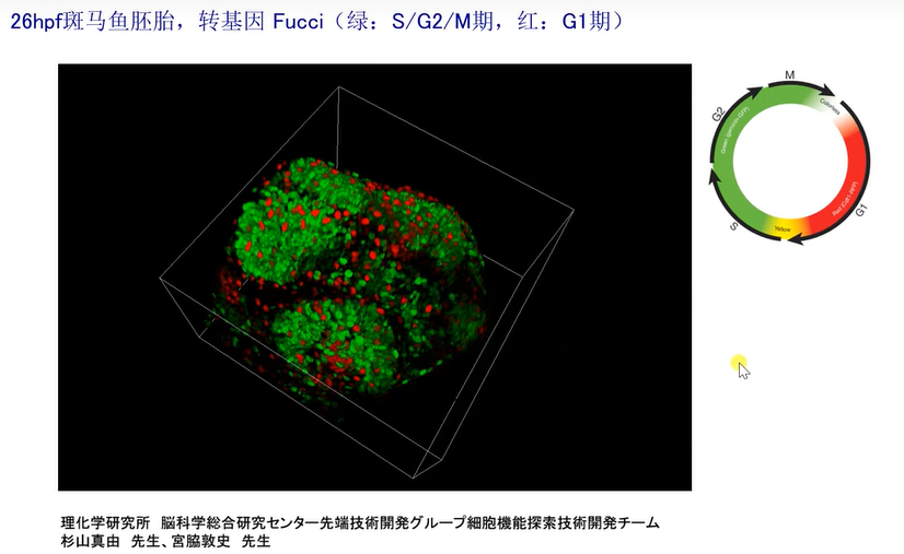
3D Reconstruction
3D Reconstruction + Multi-channel
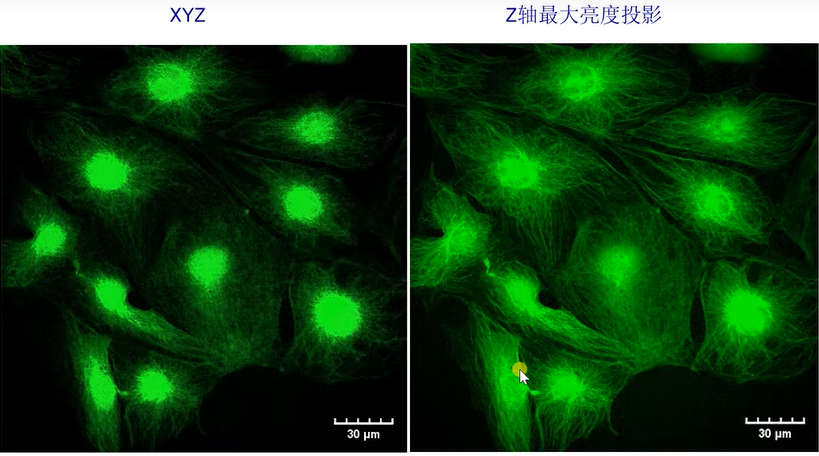
 Z-axis Maximum Brightness Projection
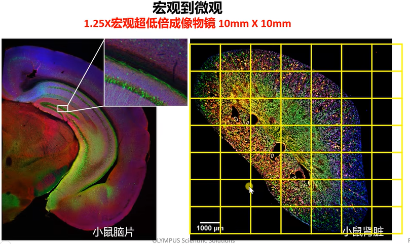
 Wide View Puzzle
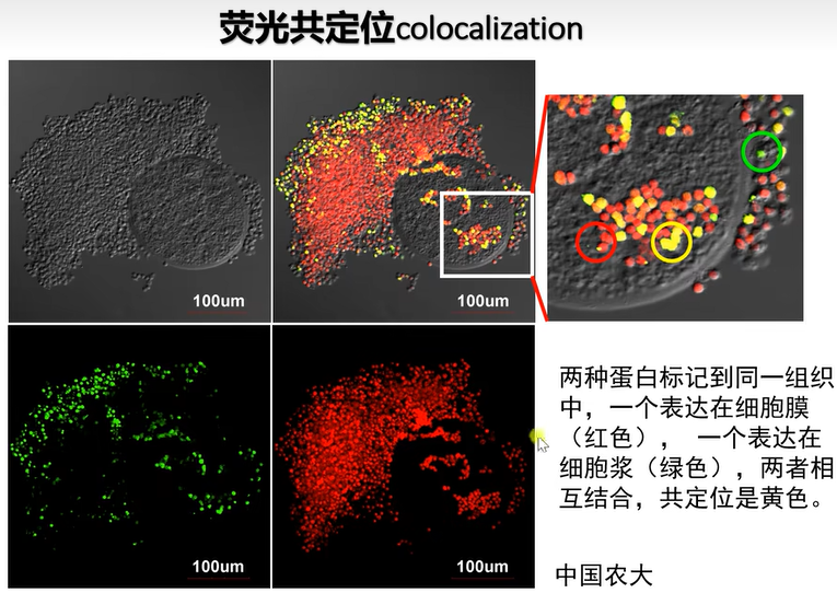
 Fluorescence Colocalization
 Time Series Imaging
Foreign Brands
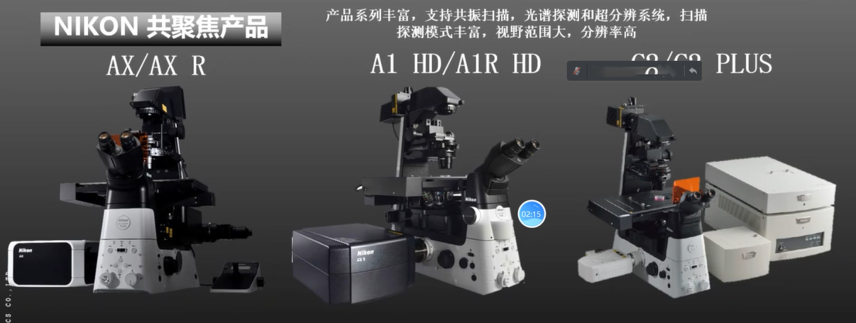 Nikon Supports Confocal
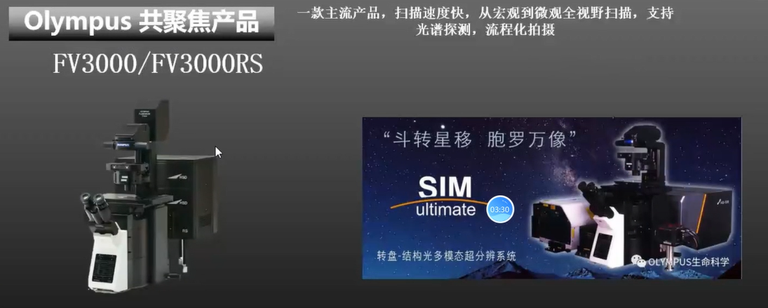 Fast Scanning Speed And Streamlined Software
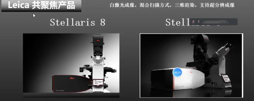 Support Super Resolution
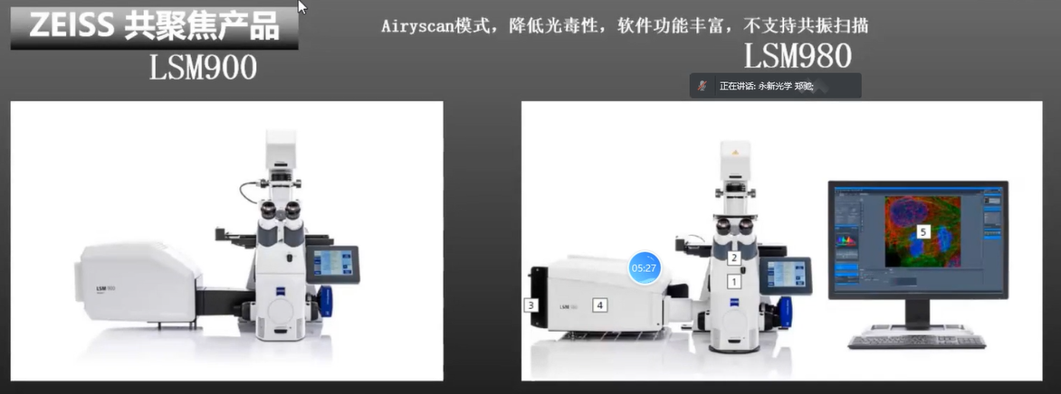 The First Of The Four, Airyscan Cellular
Chinese Model
|
|
|
|
|
|
A64.1095
|
A64.0960
|
|
A64.1095
|
A64.0960
|
|
|
Laser
|
4 Channels + AOTF
|
4 Channels
|
|
Scan
|
4096x4096
|
4096x4096
|
|
Scan Mode
|
XT, YT, XY, XYZ, XYZT
|
XY, XYZ, XYZT
|
|
View Field
|
14x14mm
|
14x14mm
|
|
Pine Hole
|
Hexagonal Zoom
|
Dia.30/40/50um
|
|
Detector
|
4 PMT + 1 DIC
|
MA PMT, GsAs PMT
|
|
Objective
|
APO
|
APO+SAPO
|
|
For any further questions or inquires about fluorescent microscopes, you are welcome to contact us at [email protected]. We will be happy to assist in selecting the Best Suit model! |
 |


 Huang Xin