 English
 Pусский
 Français
 Español
 Portugues
 Deutsch
 lingua italiana
 العربية
 Melayu
 Filipino
 Монгол хэл
 dansk
dansk Bahasa indonesia
 日本語
日本語 한국어
한국어 Türk dili
 Tiếng Việt
 svenska
 Nederlands
 Norsk språk
What is an eyepiece?
Real image: When a beam of light parallel to the optical axis passes through a convex lens, it forms scattered light. It cannot intersect at one point in the image space, only their extension lines can intersect at one point. This point is called the "virtual focus", and the plane where the focus is located is called the "virtual focus plane". The image formed by the convex lens in the object is the true focus of the light, which is called the "real image".
The main parameters of the eyepiece
Common signs for objective lens shells include:
-
WF stands for wide field of view
-
UWF stands for Extremely Wide Field of View
-
SW and SWF represent ultra-wide field of view
-
HE stands for high eye point
- CF stands for eyepiece used with CF corrected objective
-
Magnification
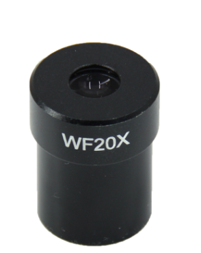
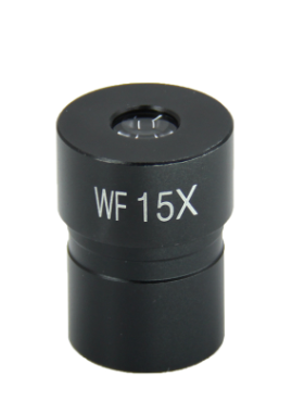
Magnification refers to the degree of visual magnification of the observed object, commonly 5x, 10x, 15x, 20x, 25x, 30x.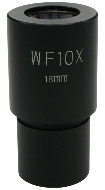
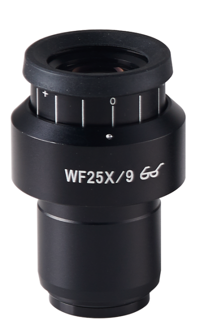
Field Number
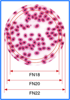
Field Number is a parameter that describes the diameter of the field of view of the microscope eyepiece . It is usually mentioned when selecting the objective lens because it needs to be used in conjunction with the objective lens. It is generally marked as FN (Field Number). It refers to the actual diameter of the sample field of view that can be observed through a specific eyepiece, usually in millimeters. The larger the field of view, the wider the sample area seen through the eyepiece. The field of view can be calculated by dividing the field of view of the eyepiece by the magnification of the objective lens. For example, if the field of view of the eyepiece is 22mm and the magnification of the objective lens is 10x, then the actual field of view diameter is 22mm.The common field of view numbers are 18mm, 20mm, 22mm, and 25mm. The larger the field of view number, the larger the field of view that can be observed, as shown in the figure below. Fields larger than 20mm can generally be called wide-angle eyepieces, while those larger than 22mm can be called ultra-wide-angle or ultra-large field of view eyepieces.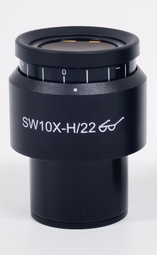
The High Eyepoint
The light transmitted from the eyepiece intersects above the eyepiece, and this intersection point is called the "eyepoint". When observing, the eye should be at the position of the eye point in order to receive all the light emitted from the eyepiece and see the maximum field of view. Otherwise, it will cause image shaking and discomfort, affecting the observation effect. The high eyepoint eyepiece is a special structure of the eyepiece. Its characteristic is that the eyepoint is far away from the eyepiece. Even if wearing glasses, the image can be observed at the eyepoint and the maximum field of view of the eyepiece can be seen. The high eyepoint eyepiece is usually marked with a glasses mark or "H".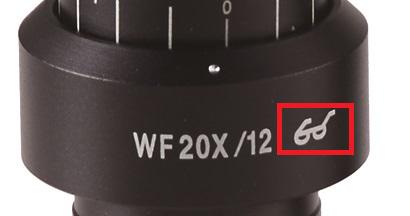
What kind of eyepiece?
Wide Field Eyepiece
Wide field eyepieces refer to flat-field eyepieces with a field of view angle of more than 50 ° and a magnification of more than 12.5x, as well as flat-field eyepieces with a field of view angle of more than 40 ° and a magnification of less than 10x. These types of eyepieces are generally called wide-angle eyepieces. The words "W" or "WF" and "WHK" are marked on the outside or surface of the eyepieces. In high-end research microscopes, some are also equipped with the letter "G". If these types of eyepieces are used in conjunction with flat-field achromatic objectives and can compensate for chromatic aberration at magnification, they are called wide-angle compensation eyepieces and marked with the letter "GB"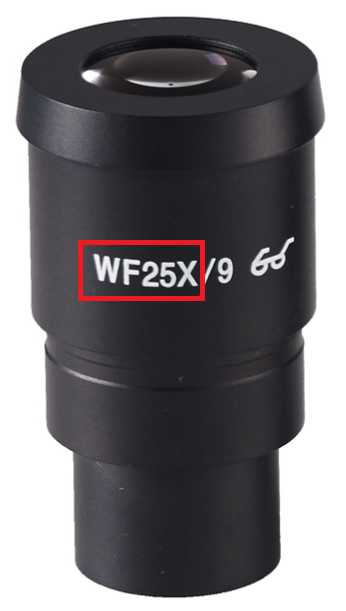
Wide Field Eyepiece with Diopter Adjustable
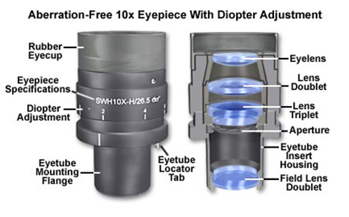
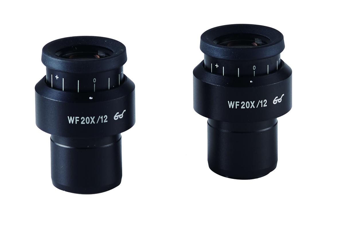
Everyone's binocular vision is different. When observing through a binocular microscope, a visual adjustment ring is generally designed on the eyepiece tube, as shown in the left figure below. Users can first observe through the left fixed eyepiece tube, adjust the focus to be clear, then observe the right eyepiece, adjust the visual adjustment ring to make the image clear at the same time, and then observe a clear image through both eyes at the same time.In addition to adjusting the visual acuity on the eyepiece tube, another way is to add a visual acuity adjustment ring to the eyepiece, as shown in the figure on the right. By adjusting the visual acuity of either side of the eyepiece, binocular observation can be ensured to be clear at the same time.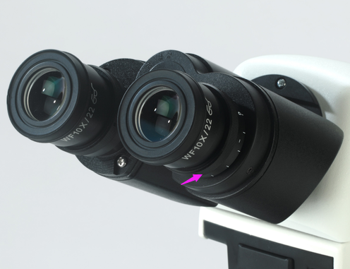
Pointer Eyepiece
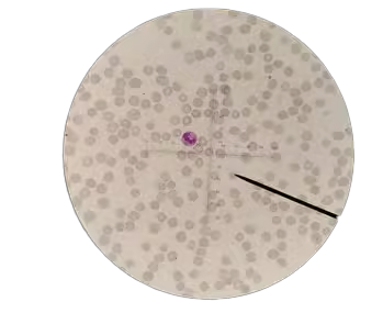
The pointer eyepiece is a part of the microscope. It has a pointer that can help us locate the position of the target on the standard plate. This pointer can rotate with the rotation of the eyepiece. When using it, you can move the stage left and right back and forth to move the object to be observed to the tip position, so that you can see the enlarged image of the object at the tip, and also determine the position of the object to be observed, which is convenient for communication with others.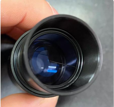
Retical Eyepiece
A micrometer eyepiece, also known as a mesh eyepiece, is a type of eyepiece made by installing a traditional micrometer tool - a micrometer ruler - on the plane of the eyepiece's field of view. It is used for microscopic measurement. The eyepiece micrometer ruler is a circular transparent glass plate with various patterns such as rulers, crosshairs, and grids etched on its surface. Its outer diameter varies according to the installation position and size of different eyepiece metal tubes, generally between 20mm and 28mm. When using a micrometer eyepiece for observation, because the ruler or crosshairs are in the same plane as the eyepiece's field of view, it is superimposed on the sample image with a clear focus. With another workbench micrometer ruler, the measurement function can be achieved by calculating the scale magnification ratio.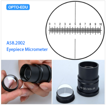
Centering Telescope
It is a special eyepiece used in conjunction with a phase contrast device. Before starting observation, inserting the centering telescope into the eyepiece tube can help adjust the center of the phase contrast condenser to the center of the optical path, ensuring good phase contrast observation effect.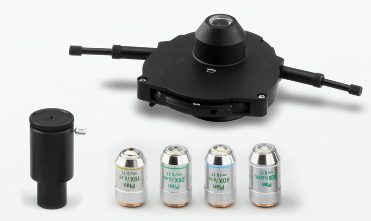
Other Eyepiece
In addition to the above several common eyepieces, there are some currently uncommon types of eyepieces, such as Huygens, Ransden, Kerner, photographic eyepieces, comparative eyepieces, projection eyepieces, etc.Huygens Eyepiece
One of the most commonly used eyepieces, named after its inventor. Structurally speaking, it consists of two plano-convex single lens eyepieces and field lenses, with the convex side facing downwards. The field diaphragm is located between the two lenses, so a crosshair or reticle cannot be installed, so it cannot be used as a micrometer eyepiece. Huygens eyepieces can effectively eliminate coma, chromatic aberration of magnification, and have very small astigmatism, but they cannot significantly reduce spherical aberration and positional chromatic aberration. Moreover, the image field is curved, protruding toward the eye end, and the field of view is very small. This eyepiece has a very low eye point, only about 3mm, which is more inconvenient to observe. It is a low-level eyepiece that is relatively simple and easy to produce, but it cannot meet the needs of high-level microscopic examination research. If an eyepiece only has a magnification marked on it, it is most likely a Huygens eyepieceRamsden Eyepiece
The Ransden eyepiece (R or SR eyepiece) is also named after the inventor. It consists of two plano-convex lenses of the same size and optical glass grade. The convex surfaces of the two lenses are opposite, and its object-square focal plane is in front of the entire eyepiece, so the field aperture is located at the lower end of the field mirror. This eyepiece can eliminate distortion and chromatic aberration , effectively reducing spherical aberration , and can be installed with crosshairs or reticle plates as micrometer eyepieces and guide eyepieces. Its eye point is higher, about 12mm, but the field of view is not large, and the plane of the field mirror is very close to the field aperture, so the dust on the field mirror can be seen directly in the field of view.Kellner Eyepiece
The eyepiece of Kellner eyepiece is made by gluing two lenses together, and is essentially an achromatic Ransden eyepiece. But it has a higher eye point than a Ransden eyepiece or a Huygens eyepiece, as well as a larger field of view. A modified version of the simple Huygens eyepiece is shown in the image above. Although these improved eyepieces perform better than simple single-lens eyepieces, they are still only suitable for low-power achromatic objectives.Photo Eyepiece, Projection Eyepiece
This kind of eyepiece is specially used for microphotography and projection. It is a negative focal length eyepiece with the eye point located in the eyepiece, so it cannot be used for observation. It is characterized by a flat field of view, which can correct the residual chromatic aberration of the objective lens, and is dedicated to microphotography. The magnification is not high, generally ranging from 2.5x to 6.7x.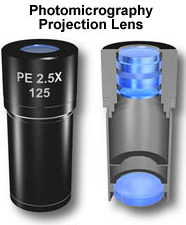
Compensation Eyepiece
Compensation eyepieces can compensate for the residual magnification and chromatic aberration of the objective lens to achieve better imaging quality eyepieces. Compensation eyepieces can be positive or negative, and should be used in conjunction with apochromatic objectives to achieve the best mirror inspection effect. Compensation eyepieces can also achieve good results if they are matched with semi-apochromatic objectives or high-power apochromatic objectives. Compensation eyepieces cannot be used with objectives with a numerical aperture below 0.65, otherwise the mirror inspection effect will be reduced. The barrel housing or end face of the compensation eyepiece is often engraved with the logo "K".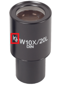
Eyepiece cleaning and maintenance
First, use a magnifier to check whether there is dust on the lens surface. If there is no magnifying glass, you can remove the eyepiece from the observation cylinder, and in turn, you can use it as magnifying glass. Adjust the angle and distance of the alignment lens surface to amplify dust or dirt. After inspection, follow the steps below to clean.-
First, blow off the dust that adheres to the surface with a blowing balloon or earball.
-
Then take a piece of mirror paper and wrap it around your finger to form a cone shape. Note that only mirror paper can be used in this step, not facial tissue, laboratory tissue or ordinary tissue. The loose coarse fibers contained in ordinary tissue can scratch the surface of the lens or stay on the lens due to breakage. When cleaning optics with a small surface area, you can fold the mirror paper into a triangular thin tip.
-
Then drop a small amount of lens cleaning solution or cleaning mixture on the tip of the mirror cleaning paper. The recommended cleaning agent is absolute ethanol and anhydrous ether mixture (mixing ratio: 70% ether, 30% ethanol), if there is no ether, anhydrous ethanol can also be used.
-
When cleaning the surface of the lens, wipe it from the center to the periphery in a rotating manner, as shown in the figure. Larger area surfaces (such as glass plates) may not be suitable for wiping using this method due to their large size. In this case, simply place the mirror cleaning paper on the surface of the wiping object, while holding the edge of the object to rotate slowly. To clean the condenser and light exit glass surfaces, you can clip a mirror cleaning paper between the middle and index fingers, and then fold it and wrap it around the index finger. When wiping the surface of the lens, press and hold the mirror cleaning paper down with your thumb. When cleaning the optical components, each mirror cleaning paper must be discarded after one use.

-
Use eyepieces or magnifying glasses to check for residual dust or dirt and make sure the lens is clean. If it is observed that the color reflected from the lens surface is not uniform, it indicates that there is dust and dirt left on the lens. In this case, another cleaning process is required until there are no contaminants on the lens.
-
After confirming that the optical components have been cleaned, immediately reinstall the cleaned components onto the microscope to ensure a clean and orderly system.
-


 Huang Xin