 English
 Pусский
 Français
 Español
 Portugues
 Deutsch
 lingua italiana
 العربية
 Melayu
 Filipino
 Монгол хэл
 dansk
dansk Bahasa indonesia
 日本語
日本語 한국어
한국어 Türk dili
 Tiếng Việt
 svenska
 Nederlands
 Norsk språk
|
What is Condenser /Iris Diagram?
What Is a Condenser on a Microscope?A condenser is a lens or system of lenses used to converge or diverge light. In microscopy, a condenser is a necessary component that provides illumination to the sample you’re viewing.
 The condenser is below or within the stage, the part of the microscope where you put the slide. It is connected to the light source. Moreover, it is positioned in a way the light shines through the stage and onto the specimen.
 The condenser on a microscope functions by collecting the light that passes through the objective lens and focusing it in one area. Since the condenser is above the light source, the collected light first passes through the observed object before reaching the condenser.
The light then passes through a hole in the bottom of the condenser and is focused on the specimen. The size of the hole in the bottom of the condenser can be adjusted to change the amount of light focused on the specimen.
 The variable-aperture diaphragm on a condenser controls the amount of light that passes through the objective lens by adjusting the size of the hole in the bottom of the condenser.
Types of Condensers on a Microscope1.Single Lens Condenser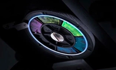 2.Abbe CondenserAn Abbe condenser is a component of a microscope. It was invented by Ernst Karl Abbe in 1870. The Abbe condenser is mounted below the stage of the microscope, and concentrates and controls the light that passes through the specimen and enters the objective.
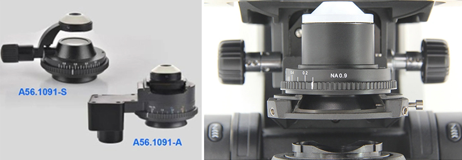
 3.Swing-Out CondenserWhen using a low-magnification objective lens (such as 4X), due to the large field of view, the light cone formed by the light source cannot fill the entire field of view, causing the edge of the field of view to be dark and only partially illuminated. To fill the field of view with illumination, the upper lens of the condenser needs to be shaken out of the optical path.
 Automatically swing-out and swing-in the top-lens element(auto Swing-out condenser of A12.1095 )
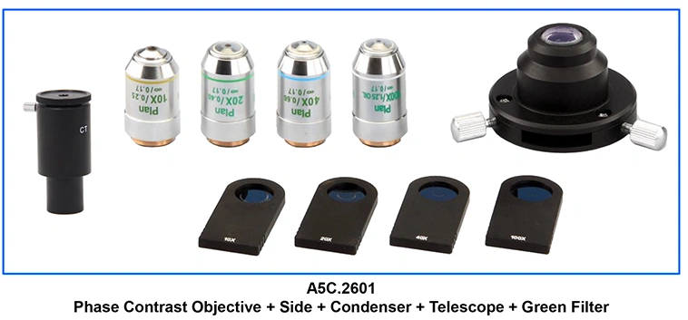
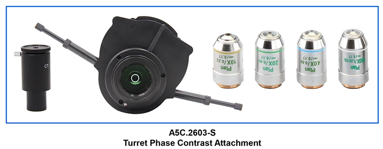
 4.Phase Contrast Condenser:In phase-contrast microscopy, the light passing through the specimen slightly shifts in phase. The shift results from different refractive indices of the specimen and the surrounding medium. The specialized condenser has a phase ring that shifts the light passing through it by a certain amount. The shift is kept in phase with the specimen, making the specimen’s details more visible.
  5.Dark Field Condenser:Darkfield microscopy is ideally used to illuminate unstained samples causing them to appear brightly lit against a dark background. A darkfield condenser scatters light and causes it to reflect off the specimen at an angle. Use darkfield microscopy to see living and unstained cells.
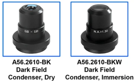 6.LWD Condenser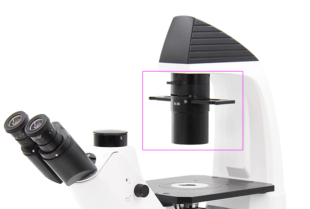 7.Universal Condenser:For upright microscope
 For inverted microscope
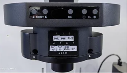 What Is A Diaphragm?A diaphragm is defined as an opaque structure with a circular opening, called aperture, at the center, which is used to control the amount of light that passes from one point to another. The diaphragm is often referred to as an aperture stop, flare stop, or field stop.
The diaphragm is located between a light source and a lens, along the optical axis of the lens system, in order for it to regulate the amount of light coming from the light source and passing through the lens. The size of the diaphragm’s aperture is what determines the amount of light.
As such, diaphragms typically feature variable sized apertures. These adjustable diaphragms are called iris diaphragms or irises.
What Is An Iris Diaphragm? From the definition of a diaphragm, an iris diaphragm is a composite type of diaphragm with adjustable or variable opening size. The name is derived from the part of the human eye aptly called the iris, which can dilate and constrict in relation to the size of the pupil.
There are many forms of iris diaphragms, the most common (and most modern) one being a set of metal or plastic blades or leaves shaped into a circle, where controlling the blades affect the size of the aperture.
How Does An Iris Diaphragm Work?As explained above, the microscope’s iris diaphragm controls the illumination and contrast of the magnified image of the specimen. The wider the diaphragm’s aperture, the higher the illumination and the lower the contrast, and vice versa.
The diaphragm can be found near the bottom of the microscope, above the light source and the condenser, and below the specimen stage. This can be controlled through a mechanical lever, or with a dial fitted on the diaphragm.
You can adjust the diaphragm by turning it clockwise to close it, or counterclockwise to open it. Only open the iris diaphragm of the microscope to a point where the light passing through barely extends beyond the microscope’s field of view.
 Iris Diaphragm Vs MagnificationThe use of a diaphragm in controlling illumination and thereby regulating the contrast is especially important in intermediate and high magnifications of a specimen under the microscope.
This is because at higher magnification levels, less light passes through, and as such, the diaphragm needs to have a wider opening to accommodate more light.
Apart from microscopy, diaphragms have plenty of uses in a variety of other optical instruments, including cameras. As with microscopy, the basic principles of a diaphragm is the same in photography.
Interestingly, however, while the goal in microscopy is to make the image as clear as possible, photography often intentionally manipulates the diaphragm to create artful out of focus spots called Bokeh. There are even specially shaped diaphragms that are designed to achieve certain imagery effects.
|
|
For any further questions or inquires about comparison forensic microscope,you are welcome to contact us at [email protected]. We will be happy to assist in selecting the Best Suit model! |
|



 Huang Xin