The Key To Micro World!

Opto-Edu (Beijing) Co., Ltd.100043Why Us (指向 公司信息, 格式不好编辑, 另起一个页面Why Us)F-1501 Wanda Plaza, No.18 Shijingshan Road, Beijing 100043, ChinaOpto-Edu (Beijing) Co., Ltd.100043Why Us (指向 公司信息, 格式不好编辑, 另起一个页面Why Us)F-1501 Wanda Plaza, No.18 Shijingshan Road, Beijing 100043, ChinaOpto-Edu (Beijing) Co., Ltd.Opto-Edu (Beijing) Co., Ltd.Opto-Edu (Beijing) Co., Ltd.Opto-Edu (Beijing) Co., Ltd.Opto-Edu (Beijing) Co., Ltd.Opto-Edu (Beijing) Co., Ltd.Opto-Edu (Beijing) Co., Ltd.Opto-Edu (Beijing) Co., Ltd.Opto-Edu (Beijing) Co., Ltd.Opto-Edu (Beijing) Co., Ltd.Opto-Edu (Beijing) Co., Ltd.Opto-Edu (Beijing) Co., Ltd.Opto-Edu (Beijing) Co., Ltd.Opto-Edu (Beijing) Co., Ltd.Opto-Edu (Beijing) Co., Ltd.Opto-Edu (Beijing) Co., Ltd.Opto-Edu (Beijing) Co., Ltd.Opto-Edu (Beijing) Co., Ltd.Opto-Edu (Beijing) Co., Ltd.Opto-Edu (Beijing) Co., Ltd.Opto-Edu (Beijing

Opto-Edu "Ultra LED" Series Fluorescence Microscope Selection and Application Guide

Opto-Edu "Ultra LED" Series Fluorescent Microscope Selection & Application Guide

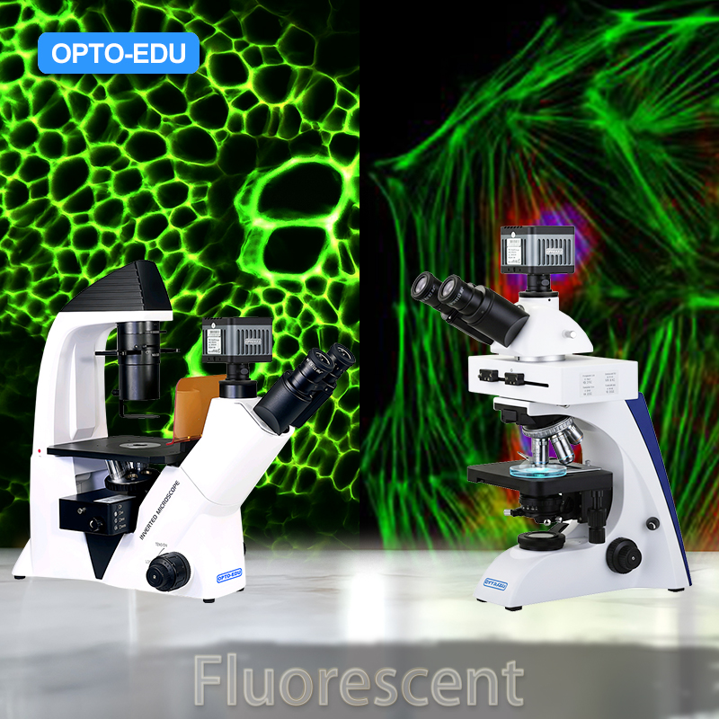
Core Recommended Models: A16.2603-UL (Upright) & A16.2614-UL (Inverted)

Technological Innovation: Why Choose the "UL" (Ultra LED) Series?

Traditional fluorescence microscopes (such as the older A16.2603 or A16.2614 models) typically employ a structure involving an "external mercury lamp housing + fiber optic/lens transmission + filter wheel." This long optical path causes significant light energy attenuation, and mercury lamps have short lifespans, generate high heat, and incur high costs.
The A16.2603-UL and A16.2614-UL adopt the revolutionary "Integrated Ultra LED" technology:
  • Integrated All-in-One Design: The high-power LED light source is directly encapsulated with the Fluorescence Filter Block.
  • Zero-Distance Illumination: The light source and filter are extremely close, shortening the optical path and resulting in virtually no light energy loss.
  • Core Advantages:
    • Brighter: The excitation light intensity is significantly boosted, offering capture capability for weakly fluorescent specimens superior to traditional mercury lamp models.
    • Purer: Complex mechanical switching mechanisms are eliminated, resulting in a blacker background and an extremely high signal-to-noise ratio.
    • More Economical: Compared to traditional models, the cost is lower, and the LED lifespan extends beyond 50,000 hours, eliminating the need for bulb replacement throughout its life.

Part I: Selection Guide (Buying Guide)

When choosing between these two models, the core decision revolves around your Sample Type (Slides vs. Culture Dishes).

Model Details and Application Scenarios

A. A16.2603-UL: Upright Fluorescence Microscope

  • Form Factor: Objective lens is above the stage; the stage is below.
  • Best Use: Pathological slices, mounted tissue samples, bacterial smears, plant sections.
  • Reason for Recommendation:
    • This is currently a highly cost-effective choice on the market. It achieves fluorescence brightness that surpasses traditionally expensive mercury lamp microscopes at a lower cost.
    • If your primary observation involves slide specimens (sections), this is the optimal choice.
    • Especially suited for applications requiring high brightness, such as tuberculosis bacillus detection, immunofluorescence (IF), and FISH (Fluorescence In Situ Hybridization).

B. A16.2614-UL: Inverted Fluorescence Microscope

  • Form Factor: Objective lens is below the stage; the stage is above.
  • Best Use: Petri Dishes, Well Plates, and live cells in culture flasks.
  • Reason for Recommendation:
    • Specifically designed for cell culture labs. Thanks to the same Ultra LED technology, it effectively avoids the weak signal issues caused by long optical paths when observing transfected live cells (e.g., GFP/RFP expression).
    • Large working space facilitates sample handling.

Comparison Table

Feature A16.2603-UL (Upright) A16.2614-UL (Inverted) Traditional Mercury Models (Old)
Sample Type Slides Dishes/Plates Slides/Dishes
Light Source Tech Ultra LED (Integrated) Ultra LED (Integrated) Mercury
Optical Efficiency ⭐⭐⭐⭐⭐ (Excellent) ⭐⭐⭐⭐⭐ (Excellent) ⭐⭐⭐ (Average)
Weak Fluorescence Excellent (Suitable for weak signal) Excellent (Suitable for weak signal) Average
Lifespan >50,000 Hours >50,000 Hours <200 Hours
Warm-up/Cool-down Instant On Instant On Requires Warm-up/Cool-down
Maintenance Cost Zero Maintenance Zero Maintenance High (Frequent bulb replacement)

Part II: Usage Instructions (Operation Manual)

Thanks to the Ultra LED technology, operating these two microscopes is much simpler than traditional models.

General Fluorescence Observation Steps

  • Sample Placement:
    • A16.2603-UL (Upright): Place the cover slip facing up.
    • A16.2614-UL (Inverted): Place the cover slip/dish bottom facing down.
  • Bright Field Locating: Turn on the transmitted light (standard illumination), find the target area at low magnification, and focus.
  • Switching to Fluorescence:
    • Turn off transmitted light (or insert the shutter) to ensure a dark background.
    • Activate the LED Module: Directly rotate/slide the turret (or slider) containing the integrated LED and filter block.
      • Select B band: Excites blue light, observe Green Fluorescence (FITC/GFP).
      • Select G band: Excites green light, observe Red Fluorescence (TRITC/RFP).
      • Select UV band (if available): Excites UV light, observe Blue Fluorescence (DAPI).
  • Brightness Adjustment: Rotate the LED brightness knob.
    • Tip: Since the UL technology is very bright, it is recommended to start adjusting from a low brightness level to protect the sample from photobleaching.

Weak Fluorescence Observation Techniques

  • Benefiting from the short optical path of the Integrated Ultra LED:
    • Adjust the brightness knob to 70%-80%.
    • Ensure the ambient light is as dark as possible (e.g., close lab curtains).
    • You will notice that the background remains perfectly black, but the target fluorescence is exceptionally bright and sharp—a feat difficult to achieve with traditional mercury lamps.

Part III: Image Capture and Large Screen Display Solution

— Recommended "Golden Partner": A59.4971 Non-cooling Fluorescent Camera

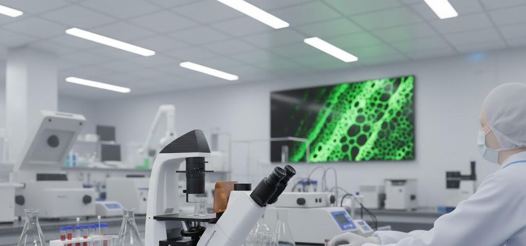
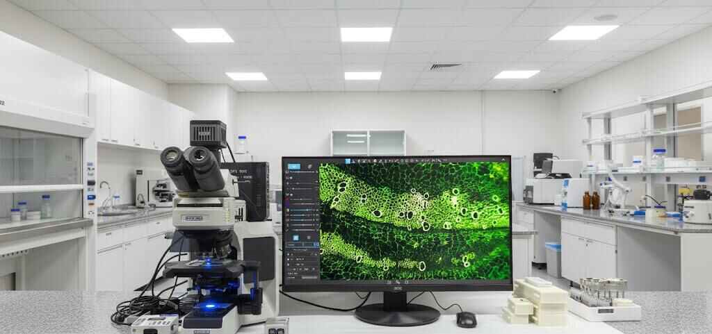
To maximize the optical performance of the Ultra LED microscopes (A16.2603-UL / A16.2614-UL) and achieve the stunning large-screen display effect like the one shown in your image, traditional cooled cameras are no longer the only option.
We highly recommend the A59.4971 Professional Non-cooling Fluorescent Camera. This camera challenges the traditional belief that "fluorescence imaging requires expensive cooled cameras," positioning it as the best accompanying solution for current fluorescence microscopes.
Technological Breakthrough: Why Choose the A59.4971 Non-cooling Camera?
  • Superior Weak Fluorescence Capture:
  • Thanks to the new generation of high-sensitivity sensor technology, the A59.4971 achieves extremely low dark current noise at room temperature. Paired with the high brightness of the Ultra LED illumination, its capability to capture weak fluorescence signals even surpasses many older generation entry-level cooled CCDs. The resulting image features a pitch-black background and clear, bright fluorescent signals.
  • Extra-Long Working Hours and Stability:
  • Traditional cooled cameras rely on fans and thermoelectric cooling, which are prone to overheating, condensation, or failure during long-term operation. The A59.4971 uses a low-power design, supporting continuous operation for extended periods without image degradation. Whether for time-lapse cell photography spanning hours or all-day pathological review, it maintains stable, high-performance output.
  • Highly Competitive Price Advantage:
  • By eliminating the expensive and complex cooling structure, while still meeting or even exceeding the performance demands of advanced research, the cost is only a fraction of traditional cooled cameras. This allows your lab to acquire a top-tier imaging system within a smaller budget.
Advanced Software Features: Making Research Easier
The A59.4971 is more than just a sensor; it features powerful integrated image processing workstation capabilities, solving two major challenges in traditional imaging:
  • Image Stitching (Large Image Mosaic):
    • Pain Point: At high magnification, the field of view is too small to see the entire tissue.
    • Solution: The A59.4971 supports real-time, automated stitching while moving the stage. You simply move the sample, and the software automatically "stitches" multiple local views into a single, high-resolution panoramic image, as easy as taking a panorama photo on a phone.
  • Depth of Field Stacking (EDF - Extended Depth of Field):
    • Pain Point: When photographing thicker tissues or cell clusters, high magnification often means one plane is in focus while others are blurry.
    • Solution: The camera supports multi-layer focus stacking. You can fine-tune the focus and capture multiple images, and the software automatically extracts the sharpest parts of each image, synthesizing a fully clear, 3D-like composite image.
Optimal Configuration for Large Screen Display
When connecting the A59.4971 to a laboratory large screen (as shown in your example images):
  • Connection Method: We recommend using the high-speed USB 3.0 interface to connect to a computer, and then using HDMI for screen mirroring.
  • Display Effect: The A59.4971's high frame rate ensures no lag and no ghosting when moving the sample on the large screen. Combined with the instant-on brightness of the Ultra LED, you can instantly display clear, highly color-accurate fluorescence images to visitors, perfectly showcasing the lab's high-end research capabilities.

Final Summary of Purchase Recommendation

The "Ultra LED Microscope + A59.4971 Camera" combination is currently the most cost-effective, lowest maintenance, and most powerful fluorescence imaging setup on the market:
  • Microscope (A16.2603-UL/A16.2614-UL): Solves the brightness and lifespan issues of the light source with integrated LED technology.
  • Camera (A59.4971): Solves the imaging cost and advanced functionality issues with high-sensitivity, non-cooling technology.
This combination will help your laboratory achieve top-tier scientific imaging results within budget.

online service

online service
We are here online to support!

Send Message

*
*
*
*
*
7
close
7

Service

Scan the QR code Close
qr code

INQUIRY LIST

Empty List

COMPARE

Empty List

Advanced Cookie Settings

Essential Cookies

These cookies enable core functionality such as security, verification of identity and network management. These cookies can’t be disabled.

Enable Marketing Cookies

These cookies are used to track advertising performance, consent to the sending of advertising-related user data to Google, and consent for the website to serve personalized advertising to provide more relevant services.

Enable Functional Cookies

These cookies collect data to remember choices users make to improve and give a more personalised experience.

Enable Analytics Cookies

These cookies help us to understand how visitors interact with our website, discover errors and provide a better overall analytics.

Essential cookies can't be disabled.