 English
 Pусский
 Français
 Español
 Portugues
 Deutsch
 lingua italiana
 العربية
 Melayu
 Filipino
 Монгол хэл
 dansk
dansk Bahasa indonesia
 日本語
日本語 한국어
한국어 Türk dili
 Tiếng Việt
 svenska
 Nederlands
 Norsk språk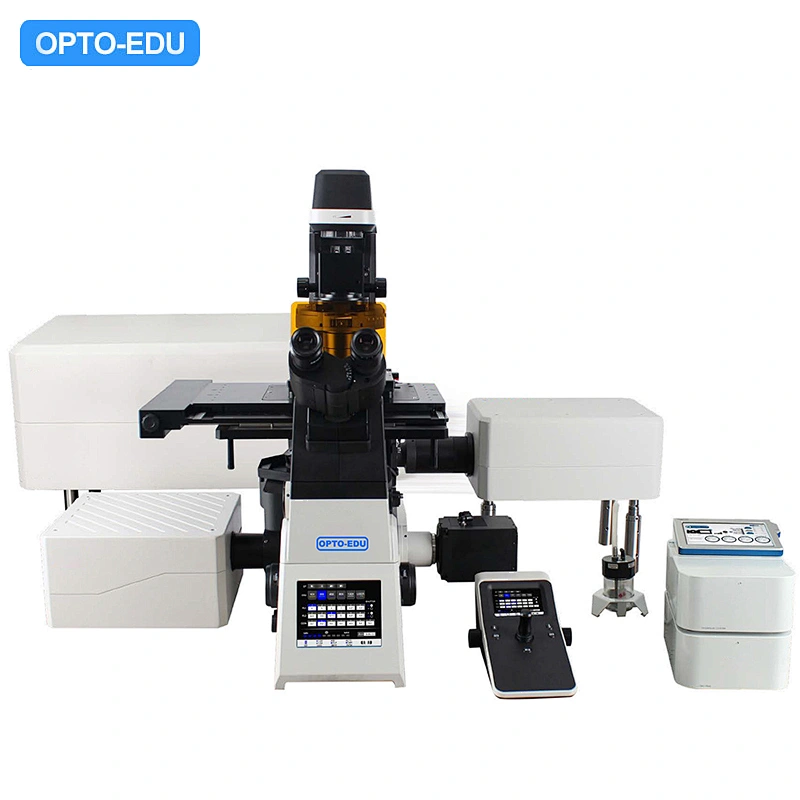
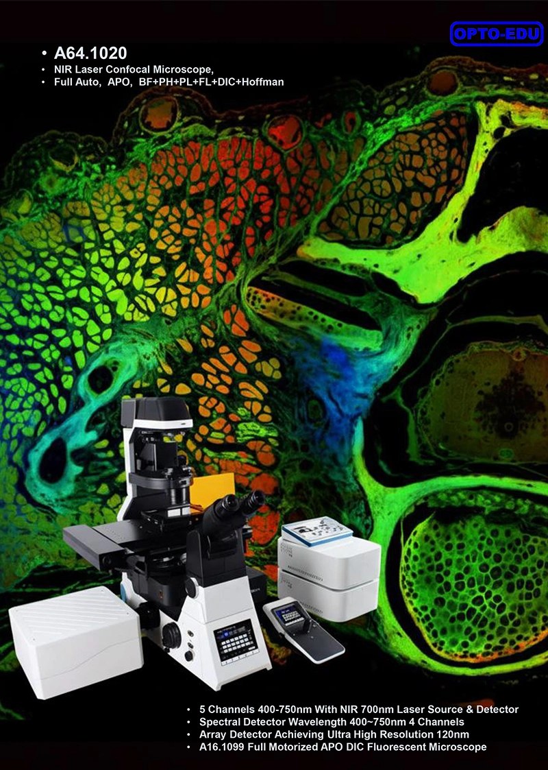
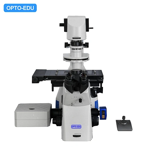
NIR Laser Confocal Microscope: Full Auto & Multi-Mode
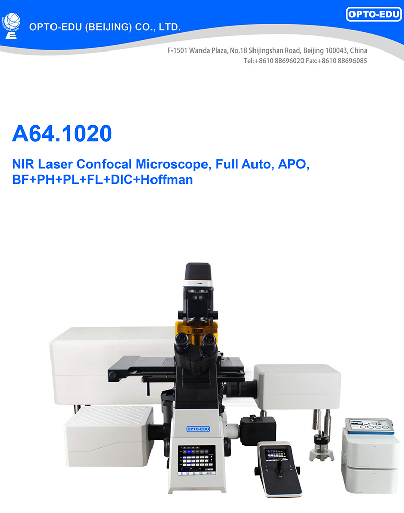
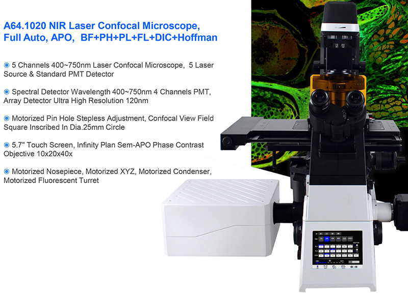
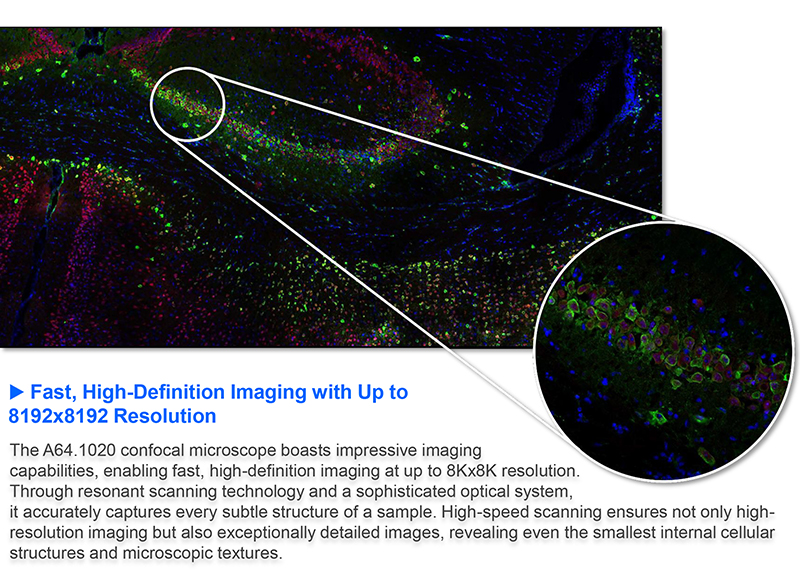
Resonance scanning, array detection, spectral imaging In life science research, analysis of tissues, organs, and model organisms is advancing at a deeper and broader level, and efficient imaging technology has become a key element in achieving breakthroughs. Acquiring images of large samples requires not only expanding the imaging range but also significantly reducing acquisition time to accurately capture internal cellular reactions. To this end, we have introduced the A64.1020 laser confocal microscope, a comprehensive evolution of the A64.1020, specifically designed to meet the demanding requirements of high-throughput, high-quality imaging in modern biological research. Inheriting the strengths of the A64.1020, the A64.1020 maintains its industry-leading 25mm field of view, enabling seamless imaging of large sample areas. Its 8192x8192 pixel scan size delivers vivid and precise images even with low-magnification microscopes. Furthermore, the A64.1020 achieves a quantum leap in key performance. Resonant scanning technology reduces acquisition time, enabling extremely high imaging speeds, and reduces phototoxicity, making it more compatible with live cell samples. The newly upgraded spatial array confocal scanner delivers low-noise, high-sensitivity spatial detection capabilities, effectively improving the signal-to-noise ratio and further enhancing resolution, allowing you to easily capture subtle structures and details within your samples. Whether capturing large-scale ultra structures or rapidly changing life processes, the A64.1020 provides precise and clear confocal imaging solutions, helping you explore uncharted territory in life sciences and uncover more of life's mysteries. |

▶High-Speed Resonant Scanning The A64.1020 utilizes resonant scanning technology, enabling extremely high-frequency scanning for rapid sample imaging. Scanning speeds are increased by nearly 20 times, shortening illumination time while maintaining image quality. This reduces photobleaching and phototoxicity, facilitating prolonged observation of sensitive samples such as living cells. |
▶More Gentle Live Cell Imaging Compared to previous point scanning technologies, resonant scanning reduces the dwell time per pixel, reducing the time the confocal laser beam remains focused on a single point. This extends the survival of living cells and significantly increases the frequency of data acquisition. This not only helps to capture high-speed dynamic events in living samples and achieve longer-term imaging observations, but also greatly improves work efficiency in fields such as drug development research, such as microplate screening. | 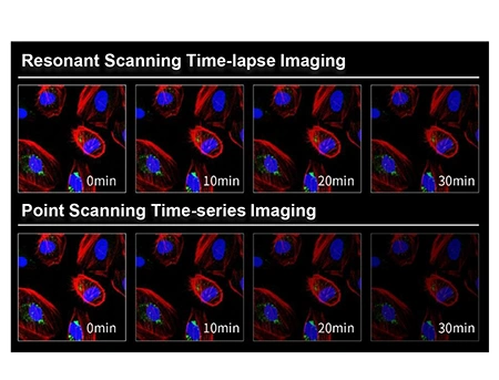 |

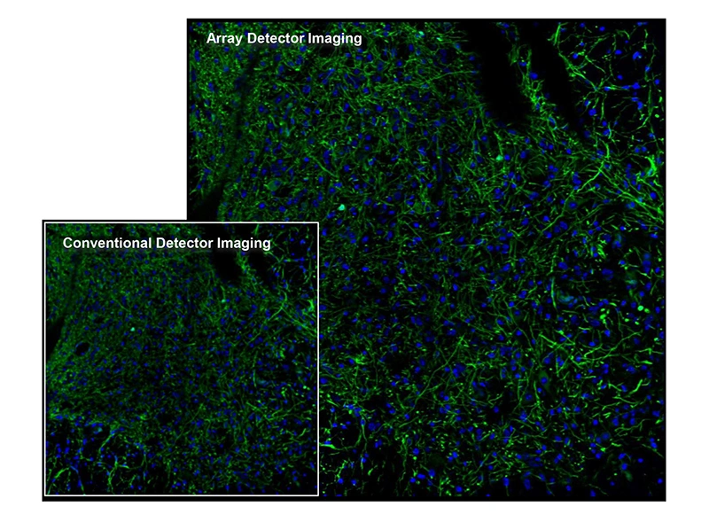
▶Low-noise, high-resolution detection The A64.1020 laser confocal microscope's array detector accurately captures extremely weak fluorescence signals, ensuring clear, detailed images. Its high resolution allows for precise resolution of sample microstructures, allowing even the smallest features to be clearly visualized. |
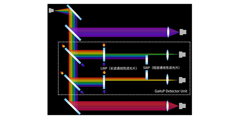
▶High Sensitivity and Accurate Spectral Decomposition The A64.1020's GaAsP photomultiplier tube (PMT) improves fluorescence acquisition efficiency, enabling users to observe weak fluorescent samples that are previously difficult to capture using traditional detection methods. The system supports up to four different spectral ranges, effectively decomposing sample-emitted fluorescence with a spectral resolution of ≤ 2nm. This effectively reduces background noise at extremely low excitation light intensities, achieving superior multicolor confocal imaging performance. | 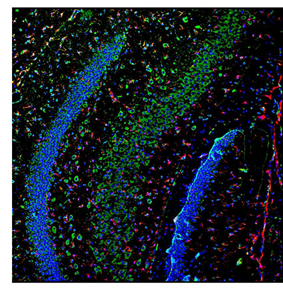 |
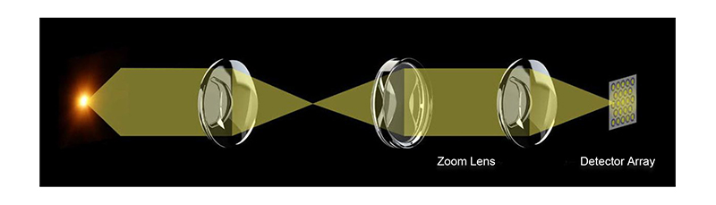
▶Space array detection technology The A64.1020's detector array, composed of multiple closely spaced SPAD detector elements, collects two-dimensional spatial information at each scan point. This two-dimensional information can be used to obtain ultrafine structures that are lost in traditional confocal detection. |
▶Spectral Unmixing System This function spectrally decomposes the fluorescence emitted by a sample, obtaining fluorescence images and spectral information at different wavelengths. This makes it possible to distinguish substances with different fluorescence emission characteristics, even if they appear similar in color under a standard optical microscope. By analyzing spectral characteristics, specific chemical components or biomolecules in a sample can be identified and authenticated, enabling qualitative analysis of the sample. |
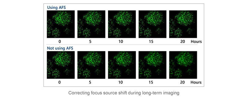
▶AFS delivers stable and reliable imaging The newly designed Adaptive Focus System (AFS) intelligently eliminates focus drift. Whether paired with high-magnification objectives with large numerical apertures or utilizing cutting-edge imaging technologies such as super-resolution, confocal imaging, and light sectioning, it consistently delivers sharp and clear images. |
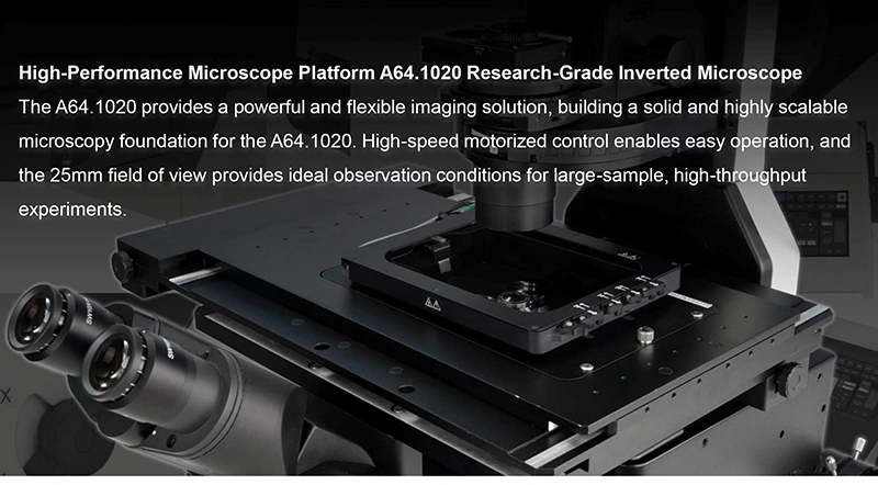
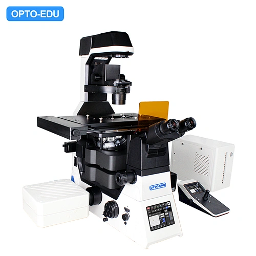
▶High-Speed Motorized Control Operation and switching speeds for objectives, filter cubes, XY stages, and observation modules have been significantly improved, creating an effortless operating environment that allows researchers to focus on daily observation and imaging. The joystick for freely controlling the stage makes using the microscope as easy as using your eyes and hands. |

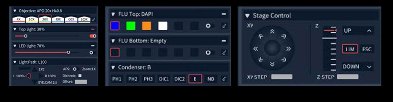
▶High Speed Hardware Control It empowers users with unprecedented convenience in operation, effortlessly digitizing management and enabling precise control over multiple electric components within the microscope, such as objective lens switching, focusing, condenser lens changing, and fluorescence module transitions. |
 |
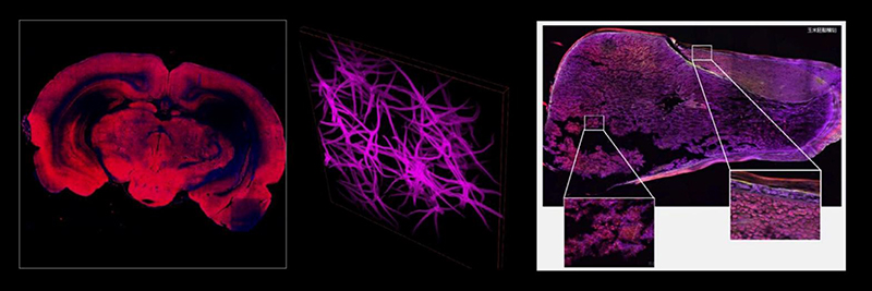
▶Multi Dimensional Imaging And Display It is capable of memorizing custom observation modes and supports the combined use of X, Y, Z, λ, and T scanning functions. Equipped with a variety of flexible shooting modes, including multi-channel fluorescence imaging, time-lapse scanning, multi-position acquisition, Z-axis stacking, and panoramic stitching. These five modes can be freely combined according to the user's actual needs, adapting to a wide range of complex and diverse experimental application scenarios. |
 |
▶Deconvolution This feature allows for deblurring of two-dimensional images. It facilitates multiple iterations of deconvolution to eliminate the out-of-focus noise components, known as speckle noise, in confocal images. Additionally, 3D deconvolution is applicable to multidimensional images, further enhancing image clarity and resolution across multiple axes. | 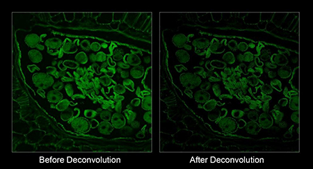 |

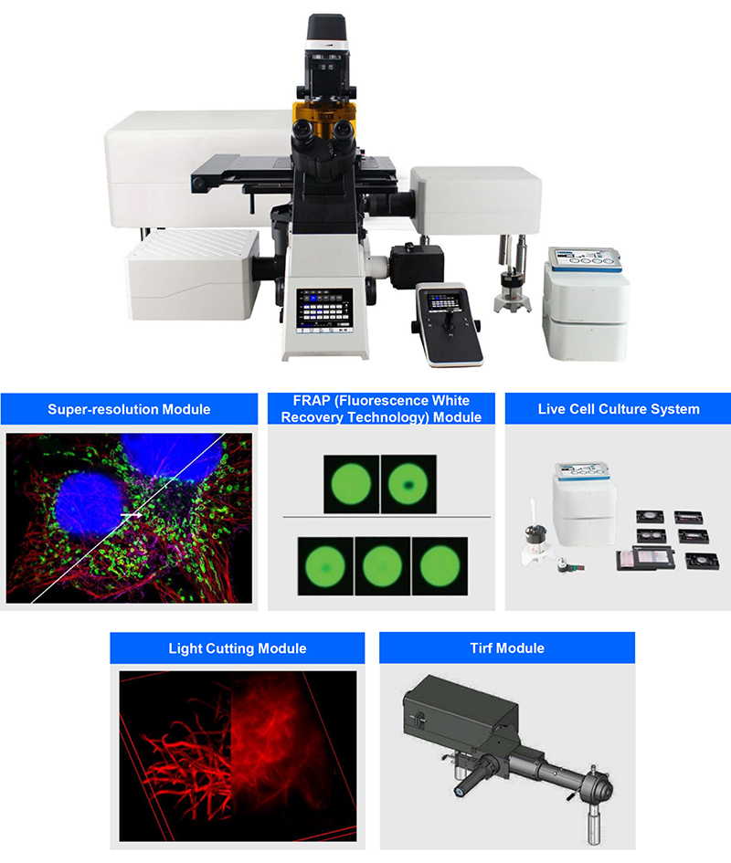
Thanks to variable structured light technology and a flexible microscope imaging platform, the A64.1020 boasts exceptional expandability. Users can easily add a variety of application accessories, such as live cell culture systems, super-resolution modules, and FRAP (fluorescence recovery after photobleaching) modules, to meet the needs of diverse application scenarios. Whether in cell biology, molecular biology, or biomedical fields, the A64.1020 provides users with comprehensive and efficient solutions. |
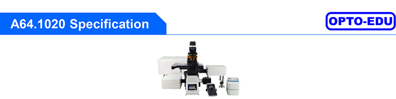
| A64.1020 Laser Confocal Microscope | Cata. No. | ||
| Confocal | 5 Channels Laser Confocal 400~750nm | ● | |
| Laser Source | 4 Laser Source With AOTF, Max Laser Power 50mW, 405nm, 488nm, 561nm, 640nm | ● | |
| 1 NIR Laser Source 730nm Optional | o | ||
| Standard Detector | Wavelength 400-750nm, 5 Channels PMT 405nm, 488nm, 561nm, 640nm | ● | |
| 730nm Optional | o | ||
| Spectral Detector | Wavelength 400-750nm, 4 Channels PMT Compatible With 4-channel GaAsP/PMT Detection, Spectral Range of 400-750nm, Adjustable Accuracy 1nm | ● | |
| Array Detector | Array-based Single-pixel Photon Counter; Achieving Ultra High Resolution 120nm | ● | |
| Scan Head | Max Pixel 8192x8192 (8K x 8K) | ● | |
| Scan Mode | Support Combine Use of X,Y,Z,λ,T Scanning Functions | ||
| Pinhole | Motorized Pinhole Stepless Adjustment, Galvanometer Scanning Mirror, | ● | |
| Field of View | Confocal Field of View: Square Inscribed In Dia. 25mm Circle | ● | |
| Depth of Image | 16 Bits | ● | |
| Computer | i7-11700/16GB*2 DDR4/1TB SSD/RTX A2000 6G/USB PCIE Card, 4x USB3.0, Win 11 | ● | |
| Monitor | 31"4K Monitor | ● | |
| Camera | 4K 8.0M 1.1"CMOS, Work With 1x C-Mount, FOV 18mm, [email protected], Max FPS46.3 | ● | |
| 24M Digital Camera, 2.7"CMOS, Work With 1.5x C-Mount (M52), 6K High Resolution, 15.3FPS@20M, Max FPS 114 | o | A55.1099-24M | |
| Port | Left Side Confocal Port With 1x Lens | ● | |
| Software | Nomis Pro X-C, Equipped With Confocal Scanning Function, Multi-dimensional Imaging, And Preset Function By Channel, Possesses Various Imaging Functions Such As Wide Field And Confocal Imaging. The Software Has Complete Control Module For The Hardware, With Built-in Basic Image Filtering And Supports Image Output In Multiple Formats. | ● | |
| Microscope | NIS60 Infinity Plan Optical System, BF, PH, Hoffman, PL, DIC, FL | ● | |
| Main Frame | Motorized Microscope Frame --5.7"Touch Screen, Show All Status, Control All Parts --Motorized Z Axis Control Working Stage Focusing --Coded Brightness Adjust Knob --F1 Button For Customized Function --Motorized Nosepiece Objective Switch --Motorized Condenser Turret Control --Motorized Layers Fluorescent Turret Control --Coded Manual Intermediate Magnification 1x/1.5x Switch Button --Coded Manual Bertrand Lens Switch & Status Detection --Motorized 4 Light Split Switch: E100/L100/R/100/E20:R80 --1x/1.5x Magnification Switch | ● | |
| Eyepiece | WF10x/22mm Eyepiece, Diopter Adjustable +/-5°, High Eyepoint | ●● | A51.1006-1022A |
| Head | 10-40° Tilt Adjustable Binocular Head, IPD 47-78mm, Eyepiece Tube Dia. 30mm | ● | A51.1099-B |
| Nosepiece | Motorized6 Holes Nosepiece, With DIC Slot, M25×0.75 | ● | A54.1099-M |
| Motorized 6 Holes Nosepiece, With Adaptive Anti-Focus Shift System (AFS) | o | A54.1099-MAFS | |
| NIS60 Infinity Plan APO Achromatic Objective | 4x, N.A.0.2, W.D.20mm, Cover Glass - | o | A52.1099-4 |
| 10x, N.A.0.45, W.D.4mm, Cover Glass 0.17mm | ● | A52.1099-10 | |
| 20x, N.A.0.75, W.D.1.1mm, Cover Glass 0.17mm | ● | A52.1099-20 | |
| 40x, N.A.0.95, W.D.019-0.21mm, Cover Glass 0.17mm | ● | A52.1099-40 | |
| 60x (Oil), N.A.1.42, W.D.0.25mm, Cover Glass 0.17mm | ● | A52.1099-100 | |
| 100x(Oil), N.A.1.45, W.D.0.13mm, Cover Glass 0.17mm | o | A52.1099-100 | |
| 100x(Oil), N.A.1.49, W.D.0.09-0.16mm, Cover Glass 0.13~0.19(23C°), 0.14~0.20(37°C) | o | A52.1099-100A | |
| Working Stage | Motorized Working Stage, Grating Type, XY Moving 130x100mm, Size 445x300mm, Max Speed 20mm/s, Moving Precision 0.1μm, Repeatability 0.5μm | ● | |
| Slide & Petri Dish Holder For Dia. 35-65mm | ● | ||
| 4~1396 Holes Plate Holder | ● | ||
| Holder For 96 Holes Petri Dish | o | A54.1099-96 | |
| Holer For For Olympus 6 Holes Petri Dish | o | A54.1099-6 | |
| Focusing | Motorized Z Axis, Optical Grating Resolution 10nm, Moving Range Up 8.5mm, Down 1.5mm, Min Step 0.02um, Repeatability 0.1um, 3 Level Focusing Knob: 2um/ 40um/ 200um Per Circle | ● | |
| Transmit Light | Kohler Illumination, With Field/Iris Diaphragm, 0~25° Titl Adjustable Arm Electrical Optical Brake Optional | ● | |
| 3W S-LED Lamp House | ● | ||
| Condenser | Motorized7 Holes Condenser Turret, --4 Small Holes Dia.37mm For Phase Contrast, Hoffman, ND Filter, --3 Big Holes Dia.39mm For DIC, ND Filters, --Up/Down Range 66mm | ● | |
| Long Working Distance Condenser NA=0.52,WD=30mm | ● | ||
| Phase Contrast | 10x-20x Phase Contrast Module | ● | |
| 40x Phase Contrast Module | ● | ||
| Coded Manual Bertrand Lens For Polarizing, Switch In/Out Light Path, Focus Adjustable | ● | ||
| DIC & Polarizing | DIC Set, Including Manual Splitter, With Optical Fiber, View Field 18mm, With DIC Prism Slide (Into Nosepiece DIC Slot), DIC Polarizer Slide, DIC Analyzer Slide | ● | A5C.1099 |
| Insert In Nosepiece DIC Slot, 10x DIC Nocaridan Prism Slide | ● | A5C.1099-N10 | |
| Insert In Nosepiece DIC Slot, 20x DIC Nocaridan Prism Slide | ● | A5C.1099-N20 | |
| Insert In Nosepiece DIC Slot, 40x DIC Nocaridan Prism Slide | ● | A5C.1099-N40 | |
| Insert In Nosepiece DIC Slot, 60x DIC Nocaridan Prism Slide | ● | A5C.1099-N60 | |
| Insert In Nosepiece DIC Slot, 100x DIC Nocaridan Prism Slide | o | A5C.1099-N100 | |
| In Condenser Turret, 10x DIC Walker Prism, | ● | A5C.1099-W10 | |
| In Condenser Turret, 20x/40x/60x DIC Walker Prism | ● | A5C.1099-W20 | |
| In Condenser Turret, 100x DIC Walker Prism | o | A5C.1099-W100 | |
| Reflect Fluorescent Light | Epi Fluorescent LED Light Source, 4 Bands 385nm, 470nm, 555nm, 630nm, With Optical Fiber & Fiber Adapter, 2 Holes Filter Insert Plate | ● | |
| Iris Diaphragm | ● | ||
| LED Light Box | ● | ||
| LED Light Controller Box | ● | ||
| Fluorsecent Turret & Filters | Motrozied 6 Holes Fluorescent Turret Disc, Motorized Shutter, With Cable | ● | |
| 2nd Layer Fluorescent Turret Set, Including Z Axis Heighten Base, Fluorescent Turnable Heighten Seat, Stage Heighten Seat | o | A5F.1099-2 | |
| FITC Filter, BP460-495, DM505, BA510-550 | ● | ||
| TRTC Filter, BP528-553, DM565, BA578-633 | ● | ||
| DAPI Filter, BP360-390, DM415, BA435-485 | ● | ||
| Middle Part | Coded Light Split Status Detection: 100/0, 0/100 | ● | |
| Mag. Switch | Coded Manual Intermediate Magnification Switch Button 1x/1.5x On Right Side | ● | |
| Light Split | Motorized Light Split Switch, Control By Front 5.7"Touch Screen: --Eyepiece 100%, --Left Photo Port 100%, --Right Photo Port 100%, --Eyepiece 20% + Right Photo Port 80% | ● | |
| Photo Port | Left Photo Port, With Built-in 1x C-Mount | ● | A55.1099-L1.0 |
| 0.7x C-Mount For Left Photo Port | o | A55.1099-L0.7 | |
| 0.5x C-Mount For Left Photo Port | o | A55.1099-L0.5 | |
| 1x C-Mount For Right Photo Port, | ● | A55.1099-R1.0 | |
| Joystick | Joystick For XYZ MotorizedControl, Touch Screen Show Objectives, Fluorscent Filters, Customized Shortcut Button | ● | |
| Tool Kit | Full Set Tool Kit For Installation | ● | |
| Dust Cover | Dust Cover For A64 Series | ● | |
| Cable | USB2.0 Cable 1.5m, Motorized Control Data Cable | ● | |
| Platform | Vibration Isolation Platform, 1000x1000mm | o | A54.1080 |


 Huang Xin