 English
 Pусский
 Français
 Español
 Portugues
 Deutsch
 lingua italiana
 العربية
 Melayu
 Filipino
 Монгол хэл
 dansk
dansk Bahasa indonesia
 日本語
日本語 한국어
한국어 Türk dili
 Tiếng Việt
 svenska
 Nederlands
 Norsk språk
How to Select OPTO-EDU Dark Field Microscope?
|
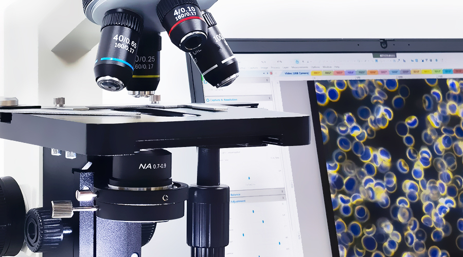
What is Dark Field Microscope |
|
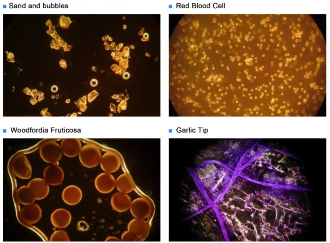
Dark field is a method designed according to the principle of Tyndall effect to observe the detected object under dark background conditions. Instead of letting the direct light enter the lens of the objective lens, let the direct light first pass through the dark field condenser and then change the path so that it enters the object obliquely, and the reflected or diffracted light on the surface enters the objective lens to form a bright image against a dark background . In the observation mode of the microscope, the dark field corresponds to the bright field, and the contrast between the bright field and the dark field is as follows |

|
What is observed in the dark field is the diffracted light image of the inspected object. It is not the object itself, so it can only see the existence and movement of the object, but cannot distinguish the fine structure of the object. But when the object to be inspected is heterogeneous and larger than 1/2 wavelength, the diffracted light at all levels enters the objective lens at the same time, and the structure of the object can be observed to some extent. Although general dark field microscopes cannot see the fine structure of objects clearly, they can distinguish the existence and movement of particles above 0.004um. This is a characteristic that ordinary microscopes (with a maximum resolution of 0.2um) do not have. The structure of cells and the movement of particles in cells, etc. |

|
Features of Dark Field Microscope ● Diffraction occurs when there is an object in the field of view, and darkness occurs when there is no object ● The background of the field of view is black, and the object is a bright color ● Due to the large incident angle, the resolution is greatly improved, and the dark field microscope can distinguish the existence and movement of 0.004um particles |

|
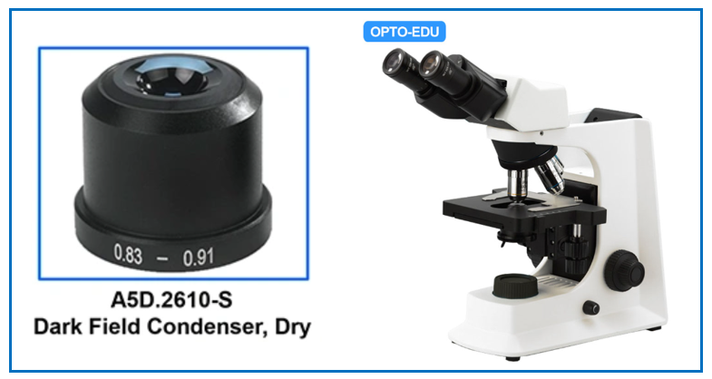
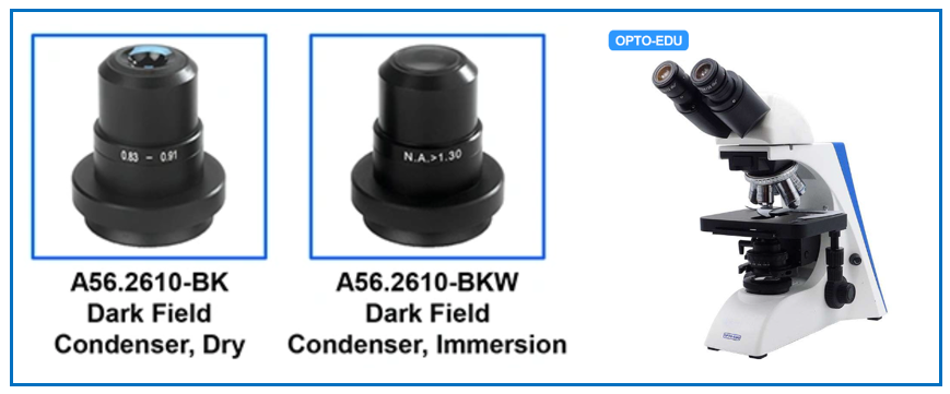
Components of Dark Field Microscope Dark Field Condenser In dark field microscopes a darkfield condenser lens must be used, which directs a cone of light away from the objective lens. To maximize the scattered light-gathering power of the objective lens, oil immersion is used and the numerical aperture (NA) of the objective lens must be less than 1.0. Objective lenses with a higher NA can be used but only if they have an adjustable diaphragm, which reduces the NA. When an opaque spider-style light stop is inserted below the completely opened aperture diaphragm, the central light rays are blocked, allowing only peripheral light rays to pass through the lenses to form an inverted oblique hollow cone of light with no change in numerical aperture. The illuminating hollow light cone is formed by refraction of light at perimeter of the lens elements, where optical correction is often poorest. Even so, this condenser will perform adequately using low magnification objectives, and produces very nice results for qualitative darkfield work. For more exacting quantitative microscopy, it is necessary to use aplanatic condensers (corrected for both chromatic and spherical aberrations), which perform much better by producing images with clearer detail and more reliable features. |

|
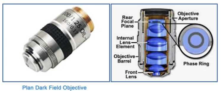
Dark Field Objective Lens Dark field objective is a special objective that is fitted with a darkened circular ring or groove (phase plate) fitted into the glass near the rear focal plane of the objective. The main feature of the dark field objective lens is that it has a hollow cone-shaped illumination beam with a large aperture. When there is no object on the sample stage, the field of view of the microscope should be dark and dull, so it is called "dark field objective". When the object to be observed is placed on the sample stage, due to the scattering, refraction and reflection of the illumination light by the object medium, the optical path of the illumination light is changed, and a part of it is scattered, refracted or reflected from the object into the microscope objective lens. |
|
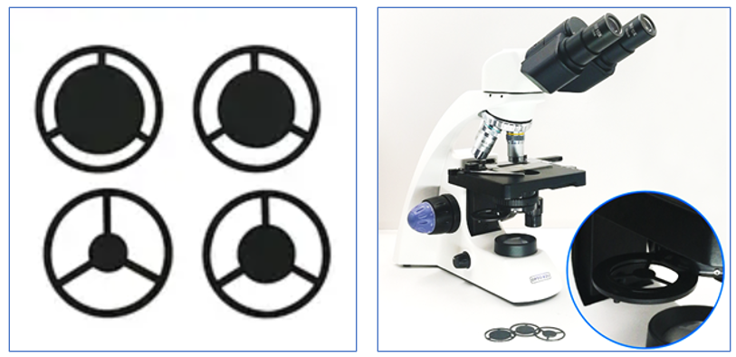
Dark Field Plates A5D.0001, a set of dark field plates, 32 mm out diameter with spider stop in the center, can be inserted into the A11.1531 microscope abbe condenser, making it as Dark Field Condenser easily. The diameter of plate can be customrized for other microscopes too. |
|
How to Use Dark Field Microscope |
|
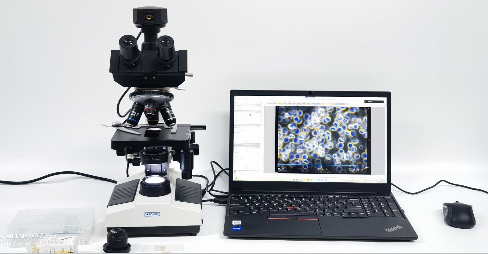
1. Replace the 10x eyepiece and 10x objective lens 2. Place the section (blood smear) 3. Replace the dark field condenser and raise it to the highest level, (if you choose a wet type, you need to add oil to the lens, and make the oil contact with the slice after raising) 4. Reduce the diaphragm of the condenser, reduce the diaphragm of the field of view, and adjust the light source to the brightest 5. Observe the field of view of the eyepiece, slowly adjust the worktable back and forth, and place the slice at the center of the optical axis 6. Slowly adjust the workbench up and down to find the object to be inspected, and adjust it until it is roughly clear 7. Slowly adjust the condenser up and down, you can see the unclear edge of the field diaphragm in the field of view 8. Adjust the optical axis Adjust the two condenser set screws to adjust the field diaphragm to the center 9. Open the condenser diaphragm and field diaphragm to the maximum 10. Turn the condenser to the lowest position, slowly turn it up, observe the field of view, you will see that the field of view is gradually covered by black, and the object under observation shows bright edges. 11. Fine-tune the height of the workbench to make the observed object the clearest |
|
Attention to the use of dark field microscope --Dark field observation requires slides and coverslips to be flawless and dust-free --Dark field observation slide cover glass is poisonous enough to meet the standard --Dry dark field condenser does not need to be refueled, it is more convenient to use, it is used for 4x10x40x --Wet dark field condenser must use cedar oil for 100x high magnification observation --During dark field observation, the numerical aperture of the objective lens is 1.0-1.25, and the numerical aperture of the objective lens with an iridescent diaphragm can be adjusted. --10.11 The two steps may need to be adjusted repeatedly to find the best effect Full Set Dark Field Microscope |
|
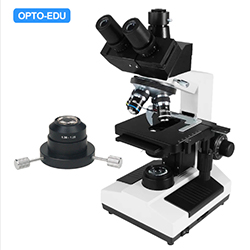
A10.1007 Dark Field Microscope ● Total Magnification 40x-1000x With Special Dark Field Attachment ● Dry Dark Field Condenser N.A.0.7~0.9 ● Immersion Dark Field Condenser N.A.1.25-1.36 ● Dark Field Objective Achromatic 100x, With Cone Diaphram ● Dark Field Objective Plan 100x, With Iris Diaphram |
|
|
|
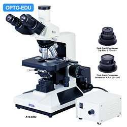
A10.0202 Dark Field Microscope, 12V50W Halogen Lamp ● Trinocular Head Light Split Switch E100:P0/E0:P100 With WF10x/20mm Eyepiece ● Quintuple Nosepiece WIth Plan Achromatic Objective 4x,10x,20x,40x,100x ● Dark Field Condenser Dry 0.77~0.91 For 20x or Lower Power Objective ● Dark Field Condenser Immersion 1.25~1.40 For 40x or Higher Power Objective ● 12V50W High Brighness Halogen Light Source Special For Dark Field View |
|
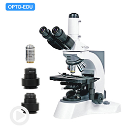
A10.1018 Professional Dark Field Microscope ● Infinity Plan Optical System WIth Objective 4x10x40x100x ● Infiniy Plan Dark Field Objective 100X With Iris Diaphram ● Dry Dark Field Condenser N.A.0.7~0.9 Work With 40x Objective ● Immersion Dark Field Condenser N.A.1.25~1.36 Work With 100x Objective ● 12V100W Kohler Illumination, Asperical Collector, Halogen Lamp |
|
|
Lab Microscope + Dark Field Attachment Most of A12. Series laboratory microscopes have optional Dark Field attachment. You can choose them freely according to your need and budget. Here are some popular models for reference: |
|
For any further questions or inquires about dark field microscopes, you are welcome to contact us at [email protected]. We will be happy to assist in selecting the Best Suit model! Written By Shirley, 2022-12 |
|



 Huang Xin