 English
 Pусский
 Français
 Español
 Portugues
 Deutsch
 lingua italiana
 العربية
 Melayu
 Filipino
 Монгол хэл
 dansk
dansk Bahasa indonesia
 日本語
日本語 한국어
한국어 Türk dili
 Tiếng Việt
 svenska
 Nederlands
 Norsk språk
What is an objective? |
|
The objective is the most important part of the microscope optical system. As part of the microscope optical system, the objective lens is mainly responsible for collecting the light emitted or reflected by the sample and focusing it to form a microscopic magnification image. The imaging quality of a microscope mainly depends on the quality of its objective lens. The design of the objective lens directly determines the key parameters such as magnification and resolution that the microscope can achieve, and is the primary standard for measuring the quality of a microscope.
|
|
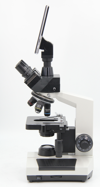
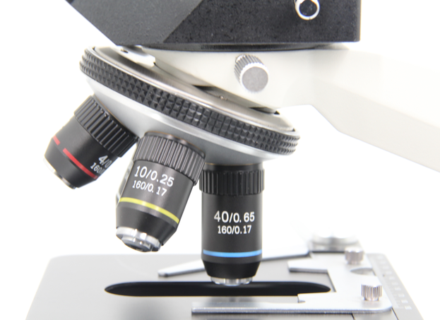
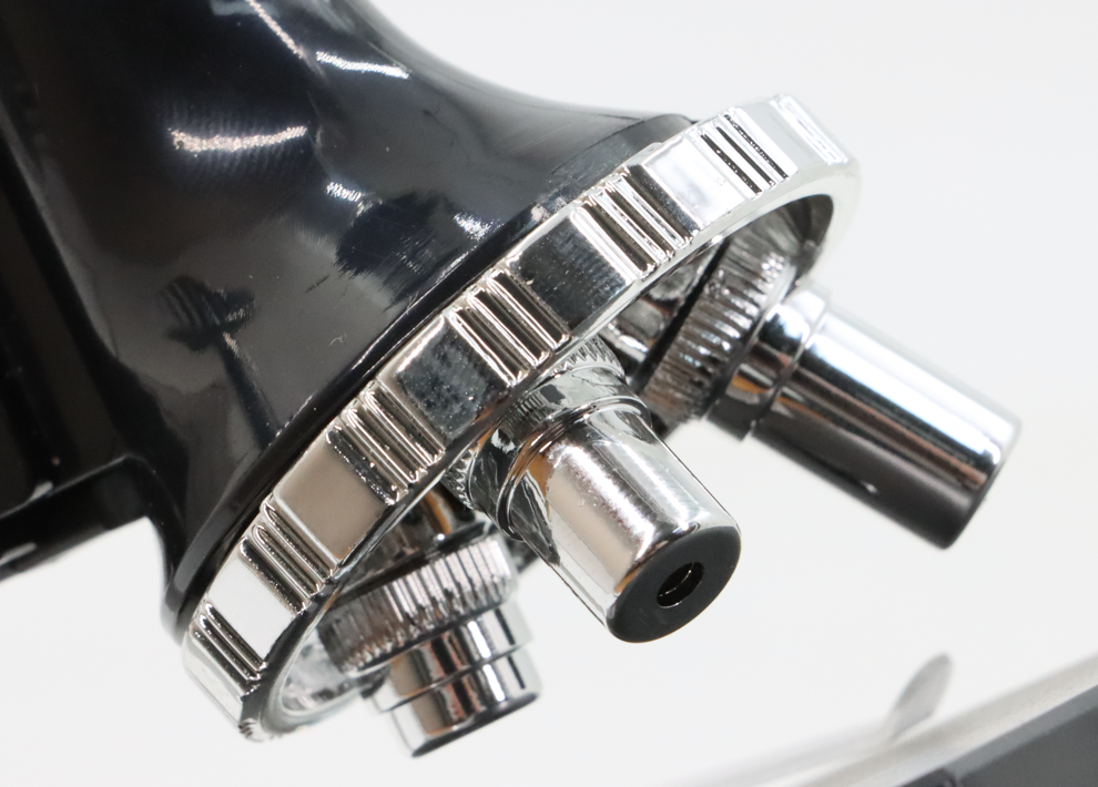
The objective lens is usually installed on the converter between the head of the microscope and the workbench, which facilitates fast and accurate movement into/out of the optical path. As shown in the figure below, The lift figure is the converter of a upright microscope, with the objective lens and workbench facing downwards to treat the observed object.
|
 |
 |
|
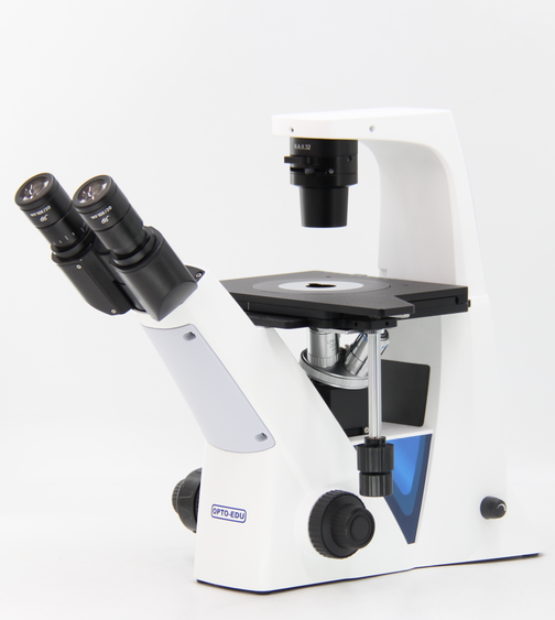
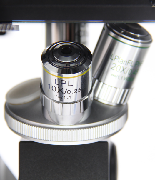
In another type of inverted microscope, the workbench and transducer positions are exchanged to form an inverted structure, and the objective lens is mounted on the transducer, facing upward, still facing the observed object.
|
 |
 |
|
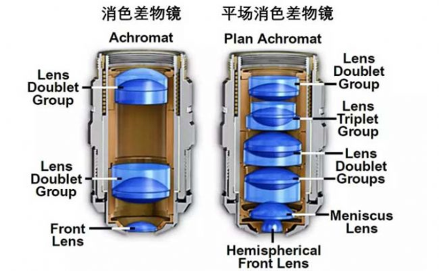
The structure of the objective lens is complex and the production is precise. It is composed of lens groups fixed at a certain distance inside the metal tube of the objective lens to eliminate aberrations. Each group of lenses is made up of one to several lenses with different materials and parameters glued together to eliminate chromatic aberrations. The front lens of the objective lens is called the "front lens", and the back lens is called the "rear lens".
|
 |
|
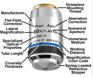
The shell of the objective lens is usually marked with some main parameters and features. The most important magnification factors, such as 10x, 40x, 100x, etc., indicate how much the objective lens can magnify the image. The numerical aperture (NA) of the objective lens is also very important because it determines the minimum resolution that the objective lens can resolve and the amount of light entering the lens. The higher the numerical aperture, the stronger the ability of the objective lens to collect light, thus providing higher resolution.
On the shell of the objective lens in the figure below, "Plan" indicates that the objective lens is a flat field objective, "APO" indicates that the objective lens is an apochromatic objective, "60x" indicates a magnification of 100x, "1.40" indicates a numerical aperture of 1.40, "Oil" indicates that the objective lens is an oil immersion objective, "∞" indicates that the objective lens is designed for infinity optics, "0.17" indicates that the coverslip thickness applicable to this objective lens is 0.17mm, "WD 0.21" indicates a working distance of 0.21mm, and "DIC H" indicates that the special optical function of this objective lens is designed for differential interference phase contrast.
|
 |
What are the main parameters of the objective lens? |
Magnification |
|
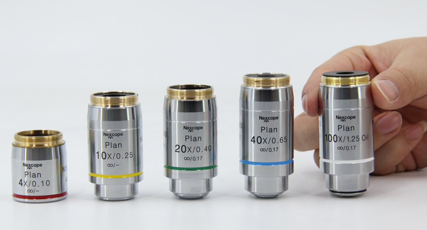
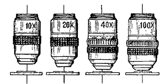
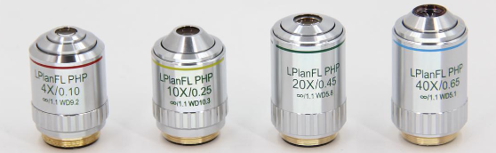
Refers to the magnification of objects observed by the objective lens, generally including 2x, 4x, 5x, 10x, 20x, 40x, 50x, 60x, 80x, 100x, etc. The following picture shows a set of infinite-distance flat-field achromatic objectives with magnifications of 4x, 10x, 20x, 40x, 100x.
|
 |
Numerical aperture: |
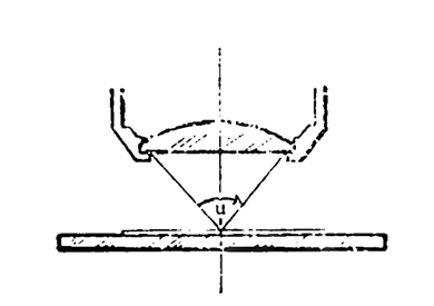 |
Numerical aperture refers to the product of the sine of the acceptance angle of the objective lens and the refractive index of the medium. It is an important parameter describing the ability of a microscope objective lens to collect light, usually marked as NA on the objective lens.
A larger acceptance angle and high refractive index medium can increase the numerical aperture, allowing the objective lens to collect more light. The larger the numerical aperture, the higher the theoretical resolution of the objective lens. When choosing an objective lens, numerical aperture is an important consideration. High-resolution applications, such as detailed imaging of cell structures or microorganisms, require objectives with high numerical apertures. For low magnification observations or when a larger depth of field is required, a lower numerical aperture is more appropriate.
|
Conjugate: |
|
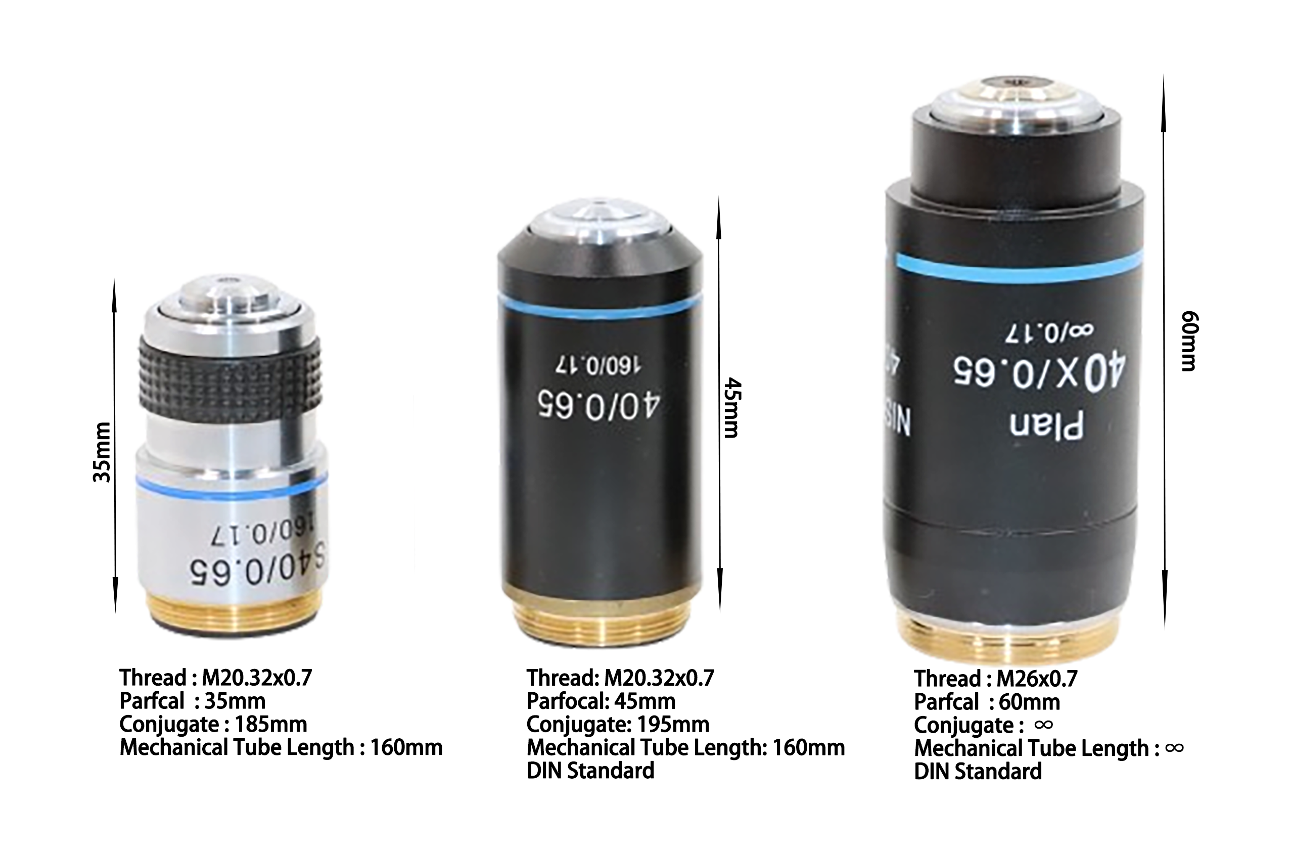
Conjugate distance refers to the distance from the object point to the image point in the microscope optical system, which is generally common 185mm, 195mm, and infinity There are three types. The 185mm conjugate distance is a common standard in early microscope designs. Microscopes of this design are usually used for standard optical observation and are suitable for education, laboratory research, and some basic industrial applications. The 195mm conjugate distance provides a slightly longer optical path for the microscope, which may allow for the insertion of more optical components and more imaging flexibility. The infinity conjugate distance objective lens refers to the objective lens not directly focusing the image at a fixed distance, but producing parallel beams of light. These beams of light are then focused by other optical components within the microscope (such as tube lenses) to form clear images. This design allows various optical components to be added between the objective lens and the eyepiece, such as reflected light sources, overview devices, filters, polarizers, etc., without affecting the focus and clarity of the image. It is very suitable for microscopes that require versatility and high flexibility.
|
 |
Parfocal and Coaxial : |
|
Zoom refers to the fact that when the objective lens of a certain magnification is used to observe the image clearly during microscopy, the imaging should also be basically clear when switching to the objective lens of other magnification, that is, the clear imaging focus of each objective lens should remain consistent. Axis This ensures that the center point of the sample deviates from the center of the field of view within a certain allowable range when the objective lens is switched. Zoom and axis usually appear together in the design of microscopes, and high-quality microscopes are all axis and focus. The combination of these two characteristics allows users to quickly switch between objectives with different magnifications without repeatedly repositioning the sample or adjusting the focus, greatly improving work efficiency and convenience of observation.
|
|
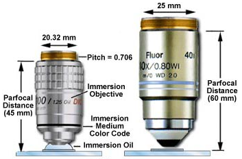
Parfocal distance is usually 35mm, 45mm and 60mm, as shown in the two examples on the right side of the figure below, which are 45mm and 60mm. Teaching microscopes have lower prices and smaller machine sizes, generally using 35mm parfocal distance objective lenses. Mid-range microscopes have larger sizes and often use 45mm parfocal distance objective lenses, while high-end microscopes have larger machine sizes and mostly use 60mm parfocal distance objective lenses. The internal space of the objective lens is larger to facilitate the design and use of more complex optical systems.
|
 |
 |
Objective thread |
| The installation thread between the objective lens and the converter generally has the following dimensions:
M20.32x0.7 (WJ4/5 "x1/36");
M25 × 0.75;
M26x0.70;
M27x0.75;
|
Mechanical tube length: |
|
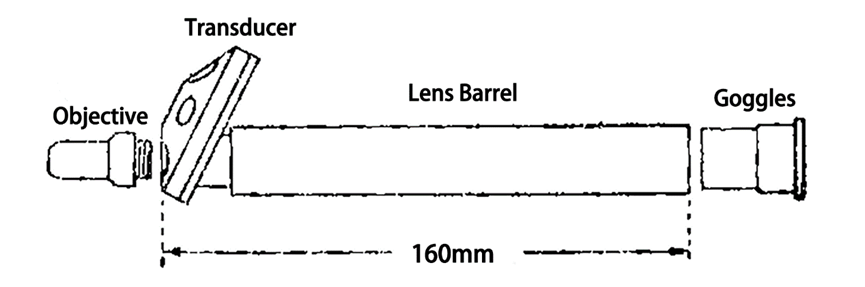
The Mechanical tube lengthof the objective lens is an important parameter in microscope design, referring to the standard length from the bottom of the objective lens to the plane where the focus is located. There are generally two types of mechanical tube lengths: infinity and standard. The standard Mechanical tube lengthis standardized as 160mm. Compared with the infinity Mechanical tube lengthsystem, their optical design is more restricted because all optical components must be correctly matched within this fixed length to achieve clear imaging. Modern microscopes are increasingly inclined to use the infinity Mechanical tube lengthsystem because it provides higher imaging quality and greater flexibility. In the infinity system, the objective lens produces parallel beams of light that can be focused at any distance, which makes it easier to insert other optical components between the objective lens and the eyepiece without affecting imaging quality.
|
 |
Working distance: |
|
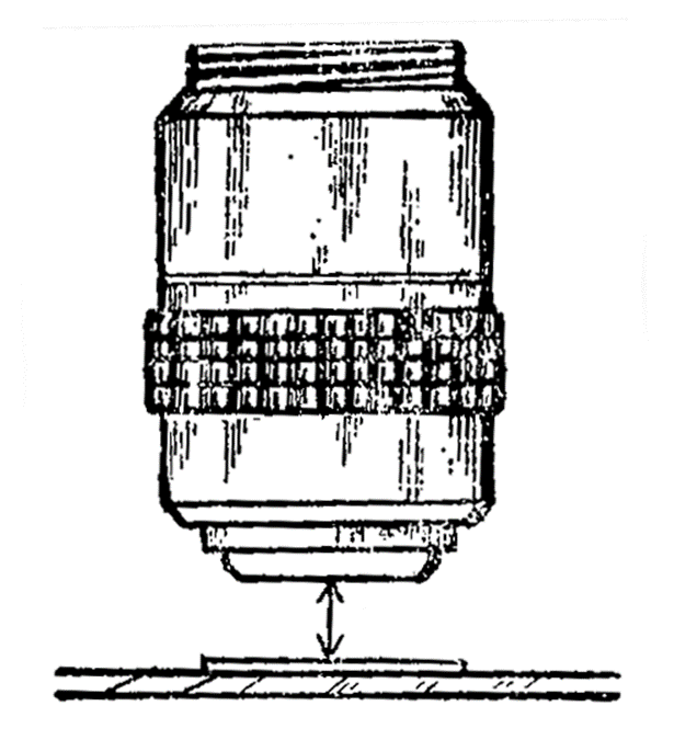
The working distance refers to the closest distance from the front end of the objective lens to the sample surface, at which the objective lens can image the sample clearly. This parameter is crucial for the convenience of experimental operations and the scope of microscopy applications. The working distance determines the available operating space between the objective lens and the sample. Long working distance objectives (LWD) provide more space for sample manipulation, processing, or tool use, suitable for metallographic microscopes that require larger operating space or inverted microscopes that need to observe through the bottom of thicker containers, which can effectively image without touching or damaging the sample. In addition, there are objectives such as ultra-long working distance ELWD and SLWD.
|
 |
Coverslip thickness : |
|
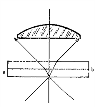
The standard thickness of a coverslip is 0.17mm, and the allowable range is 0.16 to 0.18mm. If the thickness of the coverslip is below 0.16 or above 0.18, it will bring about poor coverage, resulting in too little light or too much light entering the objective (see left image). The coverage difference of high magnification and high NA objectives is more obvious, and the thickness requirements of the coverslip are more stringent. The requirements for the thickness of the coverslip for different objectives will be indicated on the objective housing, such as 0.17. The use of coverslips of non-standard thickness may require the use of objectives with correction rings to compensate for the coverage difference.
Low magnification objectives such as 4x and 10x have a low NA value, and their imaging is not affected by the coverslip. They are labeled "-".
Long working distance objectives for metallographic microscopes, coverslips cannot be added, marked as "0"
|
 |
Field of view |
|
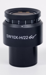
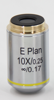
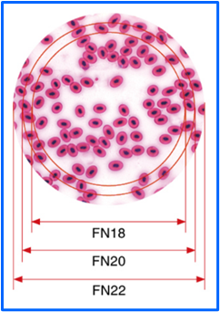
The field of view number is a parameter that describes the diameter of the field of view of a microscope eyepiece . Because it needs to be used in conjunction with the objective lens, it is usually mentioned when selecting the objective lens, and is generally labeled as FN (Field Number). It refers to the actual diameter of the sample field of view that can be observed through a particular eyepiece, usually in millimeters. The larger the field of view number, the wider the sample area seen through the eyepiece. The field of view number can be calculated by dividing the field of view number of the eyepiece by the magnification of the objective lens. For example, if the field of view number of the eyepiece is 22 mm and the magnification of the objective lens is 10 times, then the actual field of view diameter is 2.2 mm.
|
 |
 |
 |
What are the types of objective? |
|
There are many types of objective, which can be classified from different angles. According to the different correction levels of objective lenses for chromatic aberration and aberration, as well as different working distances and working media, the following are some common types on the market, which are convenient for users to choose from their own needs.
|
Toy type objective: |
|
This class of objectives generally refers to low-cost, low-quality microscope objectives with lower magnification,such as 20x or less, sufficient to observe larger samples such as salt crystals, insect wings, or plant cells. They are mostly used in toy microscopes for children or beginners.
These objectives are not designed and manufactured to the standards of professional or educational microscopes.
Toy-grade objectives are usually manufactured from less expensive materials, such as plastic or low-grade glass, which do not have the same optical properties as high-grade glass or other professional-use materials. The processing accuracy and quality control of these objectives are usually not as good as high-end objectives, which may cause image distortion or chromatic aberration. Toy-grade objectives are mainly used for educational and entertainment purposes and are suitable for children and microscope enthusiasts to conduct basic observations and experiments.
|
 |
Achromatic Objective : |
|
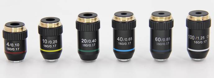
This is a commonly used microscope objective designed to correct for chromatic aberration, the problem of inconsistent focus when light of different wavelengths is refracted in a lens. This objective improves image quality by combining different types of glass to reduce the difference in focus at both ends of the spectrum. They are particularly useful for viewing fixed and stained biological samples, such as cell and tissue sections, where correction of chromatic aberration can significantly improve image quality.
|
 |
Plan Achromatic Objective : |
|
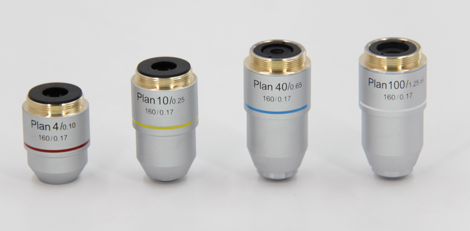
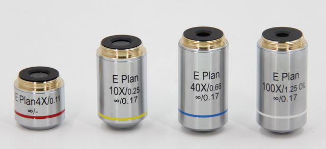
Ordinary achromatic objectives provide good imaging in the center of the field of view, but can be out of focus or distorted as they move towards the edges. A flat-field design can reduce this distortion, ensuring that the image is sharp and sharp throughout the field of view. Like ordinary achromatic objectives, flat-field achromatic objectives are also corrected for chromatic aberration, reducing the chromatic margin or blur produced by different wavelengths of light during imaging. Another key feature of this objective is better field flatness, which means that the image remains sharp and focused even at the edges of the field of view. Flat-field achromatic objectives are ideal for applications that require high accuracy and large field uniformity, such as digital imaging, precision measurement, and detailed observations that require a clear image at the edges of the field of view.
|
 |
Semi-Plan Achromatic Objective : |
|
This objective is a compromise between an achromatic objective and a flat-field achromatic objective. Semi-flat-field achromatic objectives are designed to improve out-of-focus or distortion of the field of view at the edge portion of a normal achromatic objective, providing a flatter field of view than a normal achromatic objective. Although its field of view flatness is not as good as that of a full-field achromatic objective, it is still able to provide better imaging quality from the center to the edge of a larger field of view. Semi-flat-field achromatic objectives are a cost-effective choice because they offer a compromise between price and performance. They are less expensive than full-field achromatic objectives, but offer better imaging quality than a normal achromatic objective. They are suitable for a variety of microscopy applications that require better imaging quality over a large field of view, but do not require a full-flat effect.
|
 |
Apochromatic objective : |
|
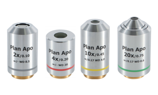
The structure of the apochromatic objective is complex. The lens is made of special glass or fluorite, fluorite and other materials, and the outer shell of the objective is marked with (APO). The apochromatic objective can not only correct the chromatic aberration of red, green and blue light, but also create images on the same focal plane to eliminate the effect of "residual chromatic aberration" (also known as secondary spectrum), and can better correct the spherical aberration of red and blue dichroic light. Due to the extremely perfect correction of various aberrations, the apochromatic objective has a larger numerical aperture than the corresponding magnification, which not only has high resolution and excellent image quality, but also has a higher effective magnification. Therefore, the performance of the apochromatic objective is very high, and it is suitable for advanced research microscopy and microphotography.
Apochromatic should be used in conjunction with compensation eyepieces during microscopy, otherwise the image quality will be degraded.
|
 |
Fluorite, Semi-Apochromatic objective : |
|
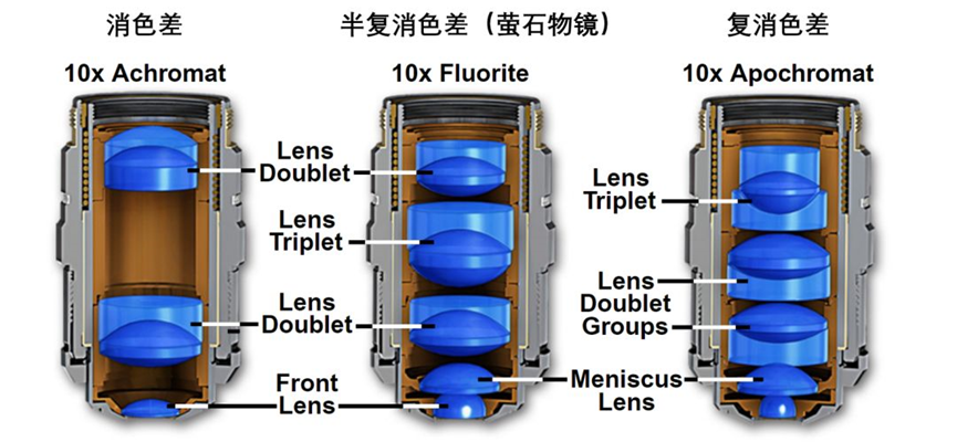
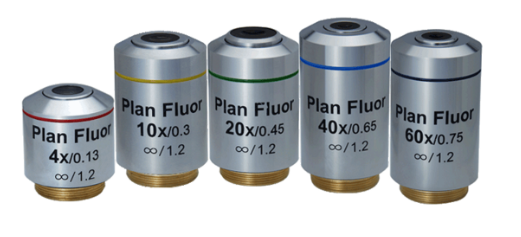
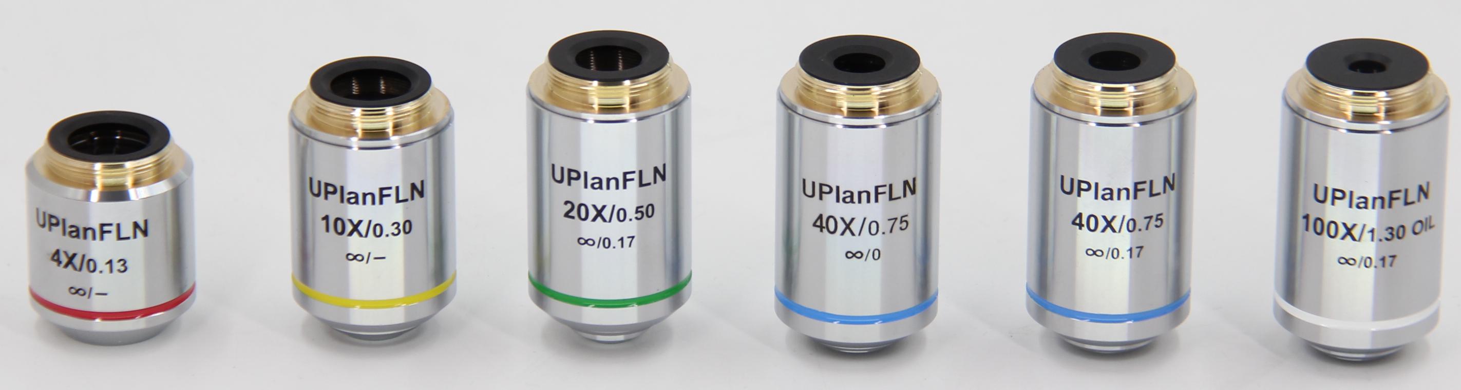
Semi-apochromatic objectives are a type of objective between achromatic and apochromatic objectives. They are designed to further reduce chromatic aberration and improve image quality, but are not as thorough as apochromatic objectives. Semi-apochromatic objectives provide better chromatic correction than standard apochromatic objectives, although they do not have the full correction capability of apochromatic objectives. This objective is designed to correct at least three different wavelengths of light to reduce chromatic aberration in the optical system. The sharpness and contrast of the image observed through the semi-apochromatic objective is higher than that of the apochromatic objective, and the chromatic edge effect is correspondingly reduced. But the overall effect is weaker than that of the apochromatic objective.
|
 |
|
Semi-apochromatic objectives offer a balanced option with a trade-off between performance and cost. They are more expensive than apochromatic objectives, but generally less expensive than apochromatic objectives, while providing image quality close to that of apochromatic objectives. Semi-apochromatic objectives offer a significant improvement over achromatic objectives in terms of color reproduction and edge sharpness. Suitable for viewing samples that require higher image clarity and reduced chromatic aberration, such as biological tissue, cells, microorganisms, and details of industrial materials.
|
 |
 |
Long Distance Objective : |
|
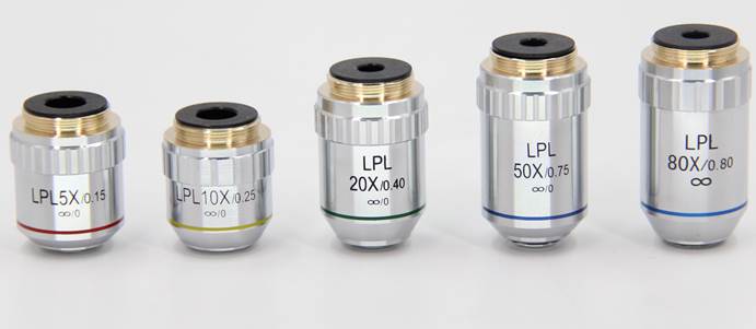
This is a microscope objective designed to operate at larger working distances, allowing the user to maintain a large space between the objective and the sample. This objective is particularly well-suited for situations where thick samples need to be viewed or manipulated over a sample. Working distance refers to the distance between the front end of the objective and the surface of the sample. Long-distance objectives provide a longer working distance than standard objectives, thus providing greater operating space. The longer working distance makes it possible to perform experimental manipulation, sample preparation, or mechanical manipulation under the microscope without interfering with the objective or the sample.
In cell culture and live cell experiments, researchers need to manipulate samples under a microscope or add reagents. Long-distance objectives are ideal for their large operating space. In addition, in fields such as materials science, metallography, and electronic engineering, long-distance objectives are used to observe large or heavy samples, such as semiconductor chips, metal parts, or to inspect surface defects.
|
 |
Oil Immersion Objective : |
|
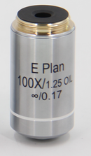
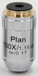
Oil immersion objective with a magnification of 90~ 100x. During microscopy, the front lens of the objective and the coverslip are usually marked with tar or non-fluorescent oil (η = about 1.515) as the medium to reduce the reflection and refraction of light, so that more light is collected by the objective lens, thereby enhancing the contrast and clarity of the image) to improve the imaging quality. In addition, glycerin (η = 1.450) and paraffin oil (η = 1.471) are sometimes used as the medium. The outer shell of the oil immersion objective is often marked with the words "Oil", "IL" or "HI".
Oil immersion objectives are widely used in cell biology, microbiology, pathology and other fields requiring high resolution imaging. They are suitable for observing small and complex structures such as cell structure, bacteria, viruses and tissue sections.
The oil-immersed objective lens must be wiped immediately after use, and cannot be left for a long time, otherwise it will damage the lens and reduce the resolution power, and the oil immersion will not be easy to wipe after drying. Wipe with a absorbent cotton ball dipped in a small amount of ether alcohol mixture (7 parts ether + 3 parts pure alcohol) to gently wipe off the oil, and then wipe it with a absorbent cotton ball or lens paper.
In contrast to oil immersion objectives, there are two other types of objectives:
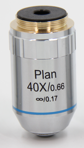
Dry objective , the objective lens between the front lens and the coverslip is air (η = 1) as the medium, the most commonly used, such as the objective lens below 40 ×, the numerical aperture is less than 1.
Water immersion objective , between the front lens of the objective lens and the coverslip is distilled water or normal saline (η = 1.333) as the medium, easy to clean. The shell is generally marked "W"
|
 |
 |
 |
|
Dry objective, oil immersion objective, water immersion objective
|
Body Vision Objective : |
|
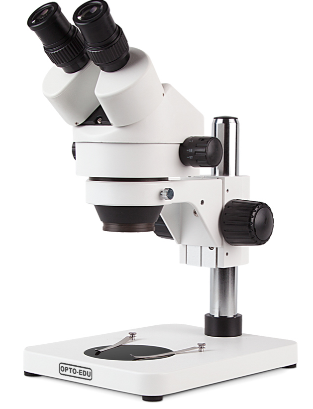
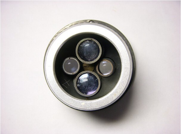
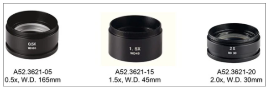
This is an objective lens designed specifically for stereomicroscopes. Unlike biological microscopes, stereomicroscopes typically have a binocular design, each with an objective lens. This arrangement can produce a stereoscopic visual effect and enhance the perception of the depth of the sample. And many stereomicroscopes today are equipped with a continuous zoom or split zoom zoom objective system, allowing the user to adjust the magnification without changing the objective lens, so as to observe different details of the sample. Stereomicroscope objectives usually provide a wide field of view, making it easy to observe the overall structure of larger samples or samples. And usually have a long working distance, providing plenty of room for the user to manipulate the sample, especially when performing surgery, assembly or fine operations.
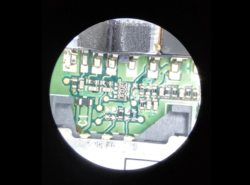
Stereomicroscopes are suitable for observing biological samples when dissection or microsurgery is required; inspecting electronic components, solder joints, mechanical parts, etc. to identify defects or perform quality control; and can also be used to examine the details of precious stones, metals, and archaeological excavations.
|
 |
 |
 |
 |
What are the special function objectives? |
A5C Phase Contrast Objective : |
|
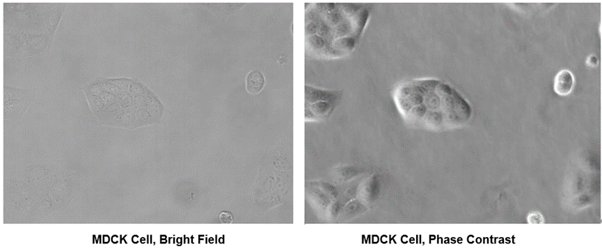
A phase contrast objective is a special type of objective used for phase contrast microscopy, characterized by the installation of a phase plate at the back focal point of the objective, which is flat. Phase contrast is a technique that allows the observation of transparent or colorless samples, such as living cells, bacteria, and other microorganisms. This technique enhances image contrast by enhancing the phase change inside the sample, making these samples, which are often difficult to observe with conventional optical methods, visible. Phase contrast microscopy relies on the phase change that occurs when light waves pass through the sample. Substances with different transparency will delay the light wave at different speeds, causing the phase change of the light wave. Phase contrast objectives, when used in conjunction with special phase contrast condensers or phase contrast ring plates, are able to separate the light passing through the sample from the direct light that has not been sampled, and cause interference in the imaging plane of the microscope, thus enhancing the contrast of the image. Phase contrast objectives are particularly suitable for the observation of transparent or translucent biological samples, such as living cells, bacteria, protozoa, and tissue cultures.
|
 |
 |
A5C DIC Differential Interference Phase Contrast Objective: |
|
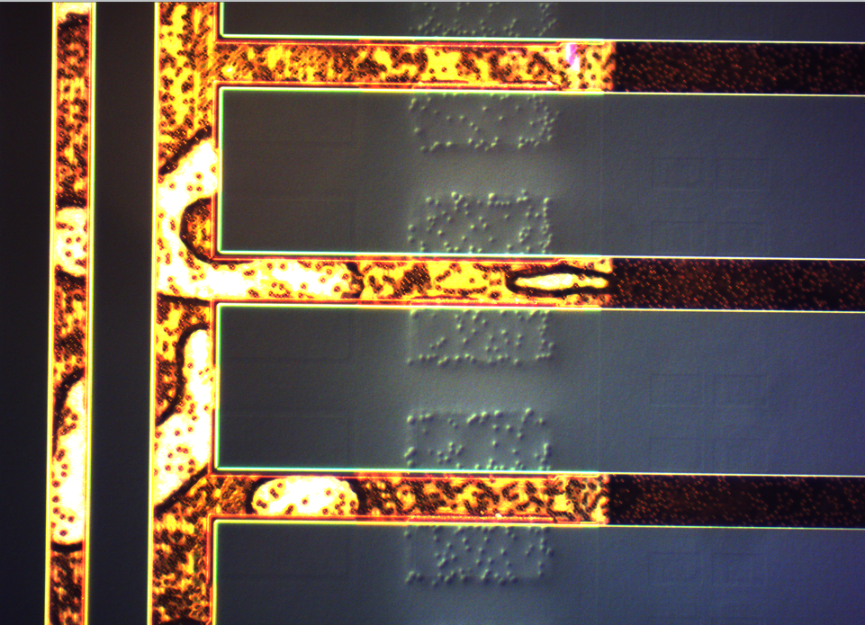
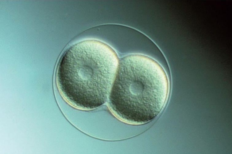
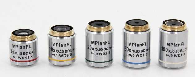
This is a special phase contrast objective used in DIC microscopy technology, which uses the interference principle of two Polarizing beams of light to enhance the contrast and edge details of the sample. The two beams of light will create a phase difference in different paths after passing through the sample. When they overlap, they will produce an interference effect, which will highlight the fine structure of the sample. The images produced by DIC microscopy have a three-dimensional sense and depth, which can show the small differences in the sample. Live cells and unstained biological tissues can be observed without the need for staining treatment of the sample.
DIC objectives are commonly used in biology, materials science, and semiconductor research. They are especially suitable for observing living cells, tissue sections, polymers, minerals, and other transparent or translucent samples. They are particularly effective for observing intracellular processes, cell-to-cell interactions, and the relationship between cells and their environment.
|
 |
 |
 |
A5C Hoffman Phase Contrast Objective: |
|
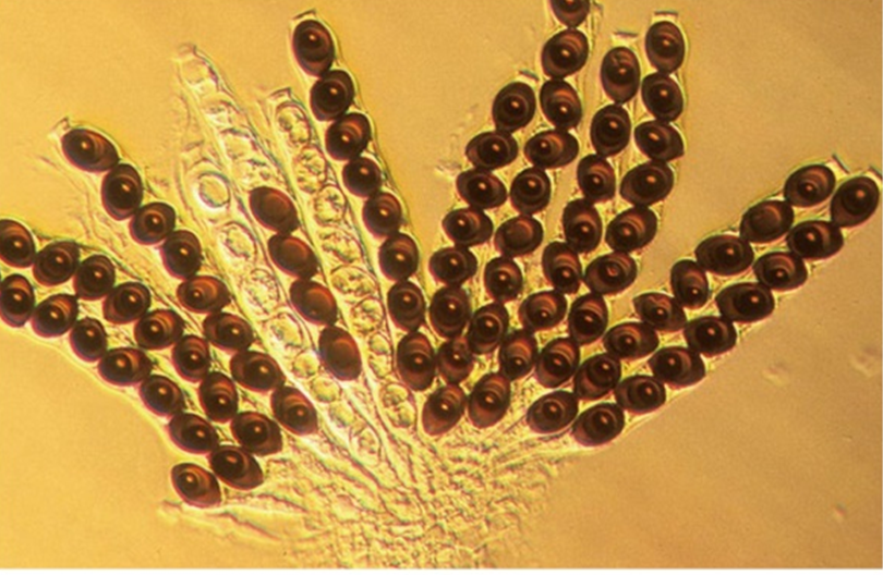
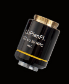
This is a special type of phase contrast objective. It is used in Hoffman modulation contrast microscopy. This objective contains specially designed optics that modulate the phase of light waves passing through the sample, converting it into a change in light intensity that enhances image contrast. When used in conjunction with a Hoffman modulator, a specific phase difference can be produced, which is used to generate high-contrast images.
Hoffmann phase contrast objectives are widely used in biological research, especially in the observation and analysis of living cells, such as cell division, migration, and the observation of intracellular organ activity.
|
 |
 |
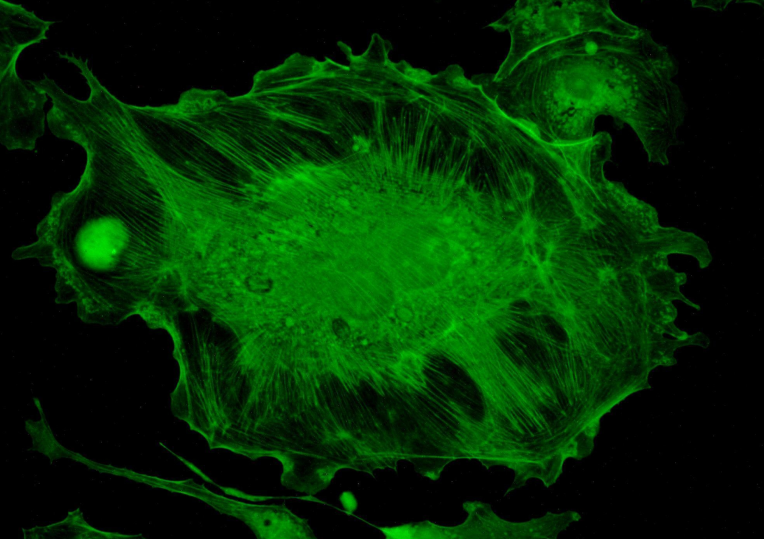
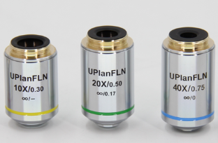
A5F Fluorescence Objective : |
|
This objective is a microscope objective designed specifically for fluorescence microscopy to view samples labeled with fluorescent dyes or labels. This type of objective has special optical properties to maximize the detection and imaging quality of fluorescent signals. Fluorescent objectives are often used in combination with specific fluorescent filters that precisely select the wavelengths of excitation and emission light, thus improving the contrast and specificity of the image. The choice of filter depends on the characteristics of the fluorescent dye used, including the wavelengths of its excitation and emission.
Fluorescent objectives are suitable for studies that require the labeling and observation of specific molecules and structures in cells or tissues.
|
 |
 |
A5D Darkfield Objective: |
|
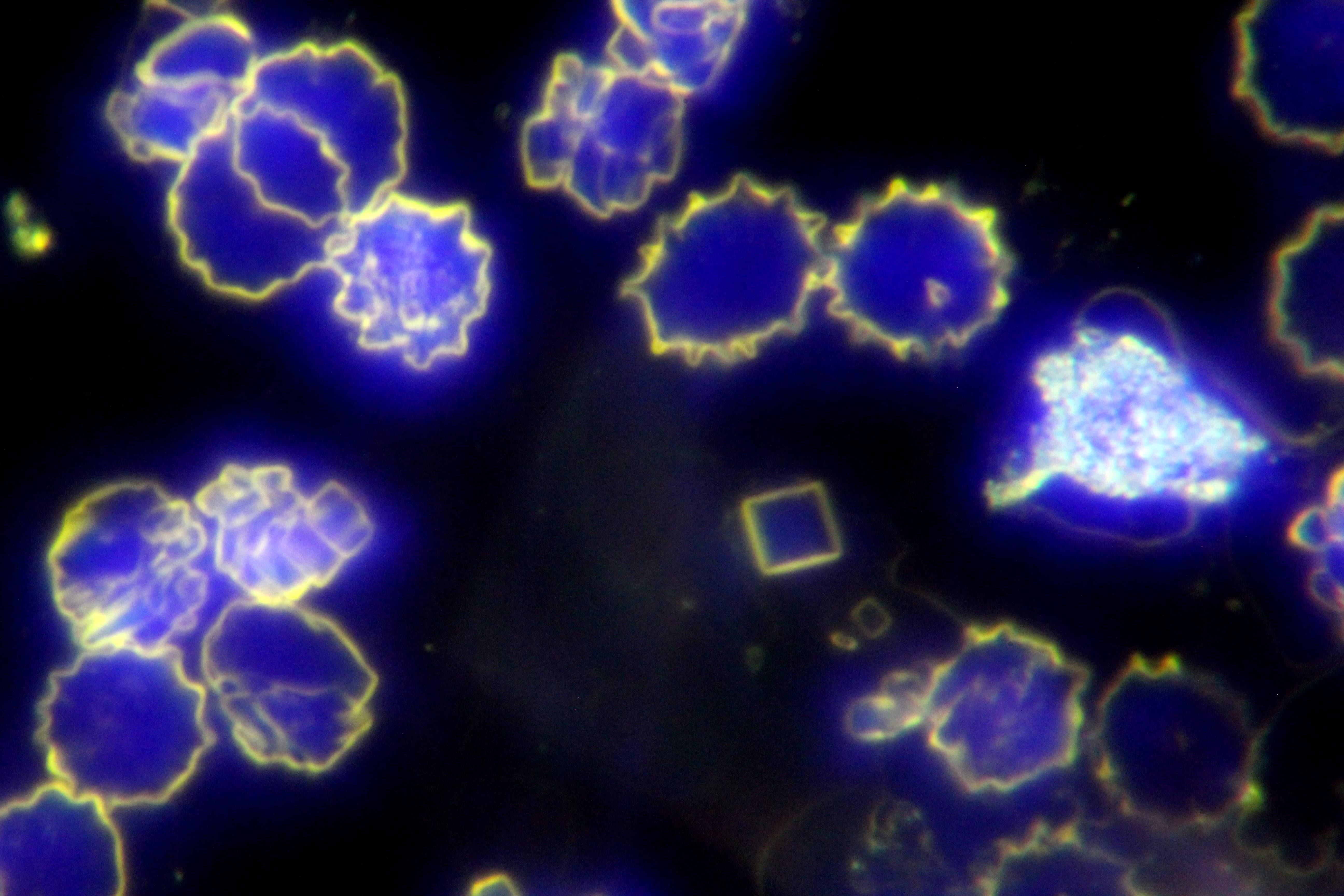
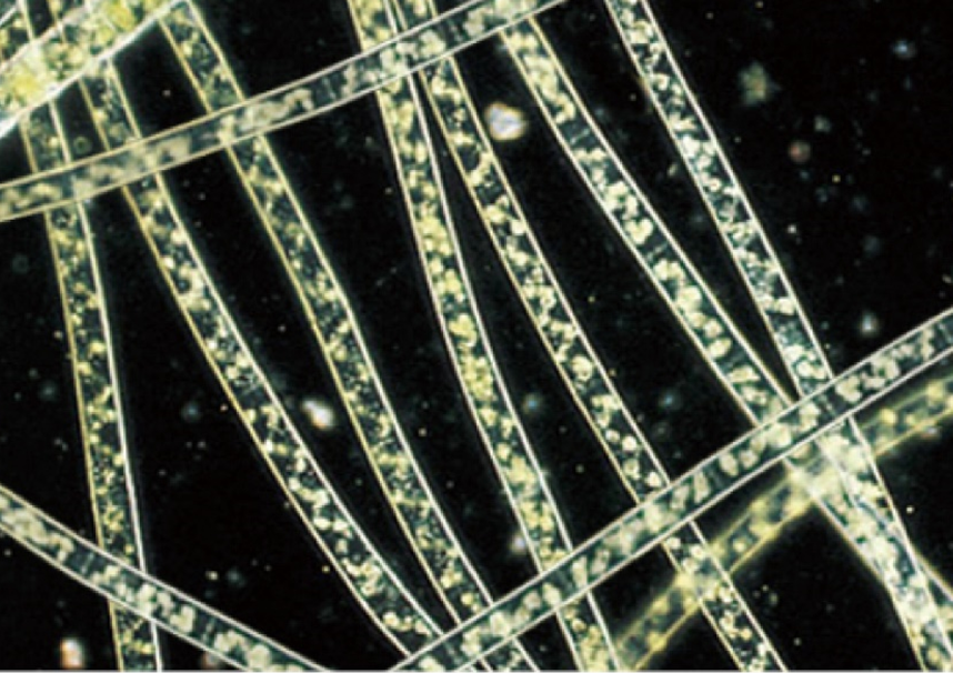
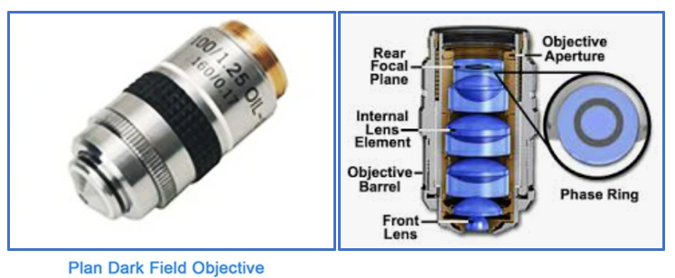
This is a specialized microscope objective used in dark-field microscopy techniques, which are able to display a bright image of the sample while the background remains dim. Dark-field microscopy utilizes lateral light to illuminate the sample, rather than direct illumination. In this configuration, only light that is scattered or refracted to the objective is captured, while direct light is blocked. This results in the sample appearing as a bright image on a dark background, as only light scattered from the sample enters the objective. In this way, the fine structure of the sample is revealed in a high-contrast form, especially for samples with less transparent and reflected light.
Darkfield microscopy is particularly valuable for studying the internal structure of cells, microbial activity, and the details of other transparent or translucent samples. It is therefore commonly used in fields such as biology, marine biology, microbiology, and materials science. It is suitable for observing living cells, bacteria, protists, and other tiny organisms or particles. However, it is important to note that dark field microscopy may not be suitable for observing samples with large thickness or strong light absorption, as these samples may block too much light, resulting in poor image quality.
|
 |
 |
 |
 |
A5D Bright and Dark Field Objective: |
|
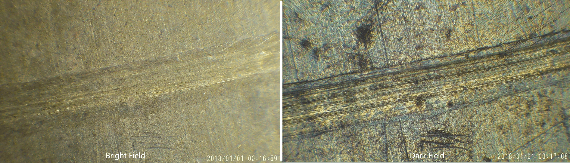
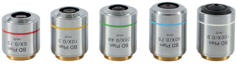
This is a special microscope objective used to achieve both darkfield and brightfield observation in microscope imaging. This objective design allows the user to switch to darkfield or brightfield imaging mode as needed to observe different characteristics of the sample. Brightfield imaging is the most common microscope observation mode, using direct light to illuminate the sample and produce an image of a dark sample on a bright background. In darkfield imaging, direct light is blocked, and only light scattered by the sample is collected. This allows the sample to appear bright on a dark background, making it particularly suitable for viewing transparent, colorless, or very fine objects. Brightfield objectives typically contain a switchable mechanism or device that allows the user to choose between darkfield and brightfield modes. This objective combines the advantages of both imaging techniques, increasing the flexibility and range of applications of the microscope.
The bright-dark field objective provides the ability to achieve multiple observation methods within a single objective, increasing the flexibility and versatility of microscopic observation. It is particularly suitable for fields requiring comprehensive analysis of samples, such as microbiology, cell biology, materials science, and environmental science.
|
 |
 |
A5P Polarizing (Stress-Free) Objective: |
|
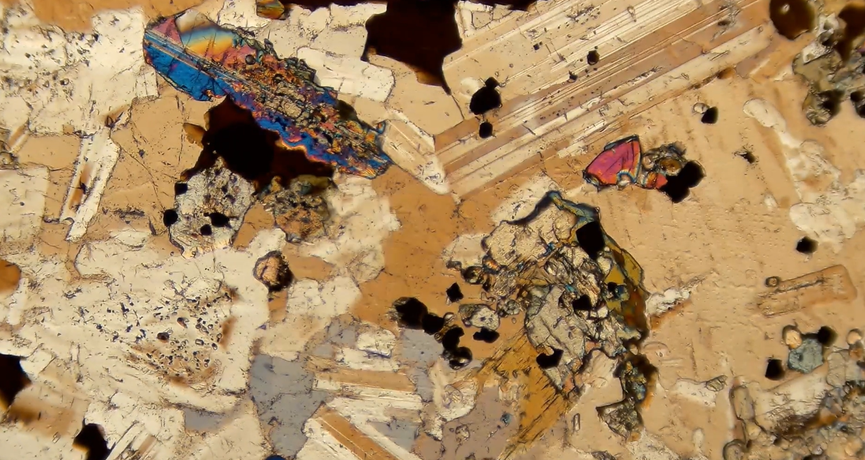
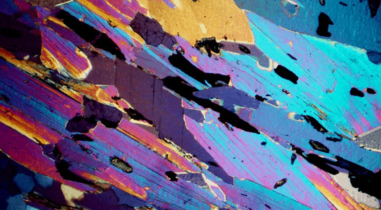
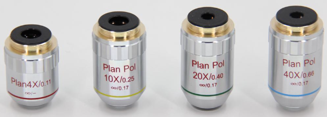
This is an objective lens designed specifically for polarizing microscopy to observe the birefringence properties of a sample. This objective is specially treated during the manufacturing process to eliminate or minimize the birefringence effect due to stress, ensuring that the observed birefringence comes from the sample itself, rather than the internal stress of the objective material. Polarizing microscopy is a microscopy technique that uses the polarization properties of light waves to study materials. When Polarizing light passes through a birefringent material, it splits into two beams along different axes depending on the internal structure of the material, resulting in a phase difference that results in a bright image.
Polarizing objectives are very useful for observing crystal structures, fibers, composites, and any samples with birefringent properties. Therefore, they are widely used in fields such as geology, mineralogy, chemistry, materials science, and biology, especially where the internal structure and properties of materials need to be studied.
|
 |
 |
 |
A5M Metallographic Objective: |
|
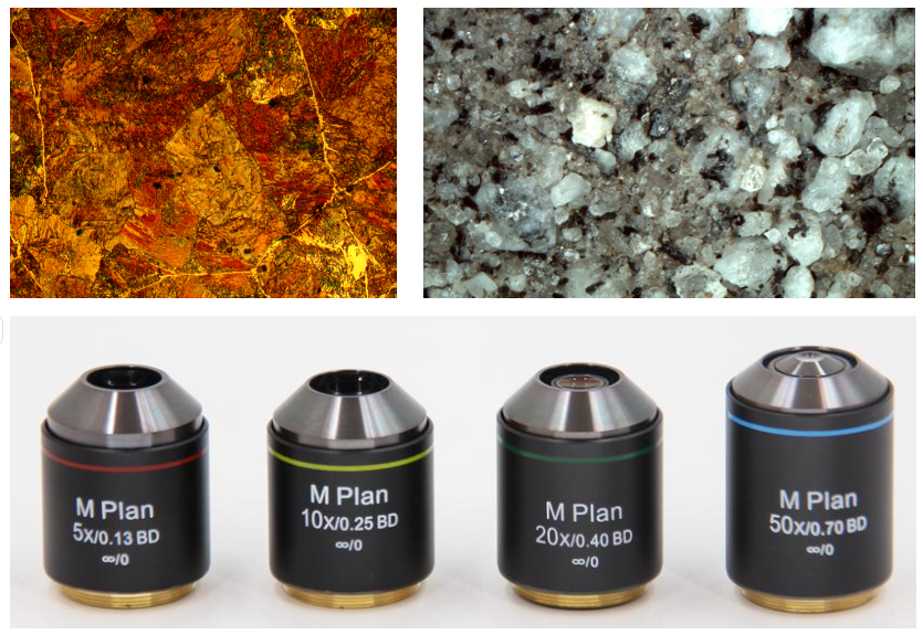
This is an objective specially designed for use in metallographic microscopes, which are mainly used to observe and analyze the microstructure of metals and alloys. Metallographic objectives are designed to illuminate with reflected light, rather than transmitted light, because the metal sample is opaque. The light source is located above the sample, and the light is directly irradiated on the surface of the sample, and the reflected light is collected by the objective lens to form an image. These objectives usually have the characteristics of high resolution, long working distance, and no cover glass, which can reveal the microstructure of metals and alloys without imposing major restrictions on the observed object in terms of size.
Metallographic objectives enable users to study the microstructure of metals in detail, including composition distribution, crystal orientation, and defect types. Widely used in materials science and engineering to study the microstructure of metals and alloys, in industrial manufacturing, metallographic microscopes are used to inspect the quality of metal parts and materials
|
 |
Other special objectives |
|
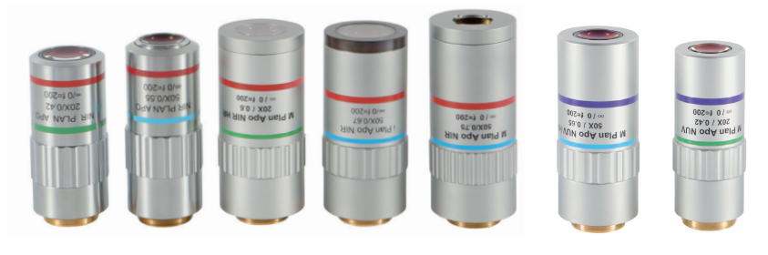
In addition to the objectives described in detail above, there are also some special objectives that are relatively less used, such as near-infrared objectives, near-ultraviolet objectives, white light interference objectives, etc. Each type of objective has its advantages and disadvantages, and the choice of objective will depend on the specific application and user needs
|
 |
Common abbreviations for objective lenses |
|
Optical Corrections These are usually listed as Achro and Achromat (achromatic), as Fl, Fluar,Fluor, Neofluar, or Fluotar (fluorite) for better spherical and chromatic corrections, and as Apo (apochromatic) for the highest degree of correction for spherical and chromatic aberrations. Field curvature corrections are abbreviated Plan, Pl, EF, Achroplan, Plan Apo, or Plano. Other common abbreviations are ICS (infinity corrected system) and UIS (universal infinity system), N and NPL (normal field of view plan), Ultrafluar (fluorite objective with glass that is transparent down to 250 nanometers), and CF and CFI (chrome-free; chrome-free infinity). The objective in the illustration (Figure 1) is a plan apochromat that enjoys the highest degree of optical correction. See Table 1 for a complete list of abbreviations often found inscribed on objective barrels.
|
| ABBREVIATION | TYPE |
| Achro, Achromat | Achromatic aberration correction |
| Fluor, Fl, Fluar, Neofluar, Fluotar | Fluorite aberration correction |
| Apo | Apochromatic aberration correction |
| Plan, Pl, Achroplan, Plano | Flat Field optical correction |
| EF, Acroplan | Extended Field (field of view less than Plan) |
| N, NPL | Normal field of view plan |
| Plan Apo | Apochromatic and Flat Field correction |
| UPLAN | Universal Plan (Brightfield, Darkfield, DIC, and Polarizing Light) |
| LU | Luminous Universal (Brightfield, Darkfield, DIC, and Polarizing Light) |
| L, LL, LD, LWD | Long Working Distance |
| ELWD | Extra-Long Working Distance |
| SLWD | Super-Long Working Distance |
| ULWD | Ultra-Long Working Distance |
| Corr, W/Corr, CR | Correction Collar 校正环 |
| I, Iris, W/Iris | Adjustable numerical aperture (with iris diaphragm) |
| Oil, Oel | Oil Immersion |
| Water, WI, Wasser | Water Immersion |
| HI | Homogeneous Immersion |
| Gly | Glycerin Immersion |
| DIC, NIC | Differential or Nomarski Interference Contrast |
| CF, CFI | Chrome-Free, Chrome-Free Infinity-Corrected (Nikon) |
| ICS | Infinity Color-Corrected System (Zeiss) |
| RMS | Royal Microscopical Society objective thread size |
| M25, M32 | Metric 25-mm objective thread; Metric 32-mm objective thread |
| Phase, PHACO, PC | Phase Contrast |
| Ph 1, 2, 3, etc. | Phase Condenser Annulus 1, 2, 3, etc. |
| DL, DLL, DM, BM | Phase Contrast: Dark Low, Dark Low Low, Dark medium, Bright Medium |
| PL, PLL | Phase Contrast: Positive Low, Positive Low Low |
| PM, PH | Phase Contrast: Positive Medium, Positive High Contrast (Regions with higher refractive index appear darker.) |
| NL, NM, NH | Phase Contrast: Negative Low, Negative Medium, Negative High Contrast (Regions with higher refractive index appear lighter.) |
| P, Po, Pol, SF | Strain-Free, Low Birefringence, for Polarizing Light |
| U, UV, Universal | UV transmitting (down to approximately 340 nm) for UV-excited epifluorescence |
| M | Metallographic (no coverslip) |
| NC, NCG, - | No Coverslip |
| EPI | Oblique or Epi illumination |
| TL | Transmitted Light |
| BD, HD, B/D | Bright or Dark Field (Hell, Dunkel) |
| D | Darkfield |
 |


 Huang Xin