 English
 Pусский
 Français
 Español
 Portugues
 Deutsch
 lingua italiana
 العربية
 Melayu
 Filipino
 Монгол хэл
 dansk
dansk Bahasa indonesia
 日本語
日本語 한국어
한국어 Türk dili
 Tiếng Việt
 svenska
 Nederlands
 Norsk språk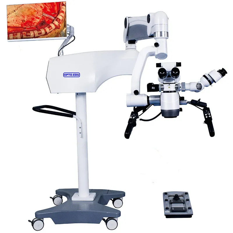

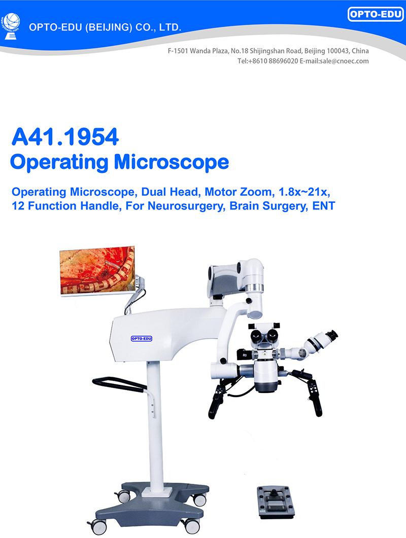
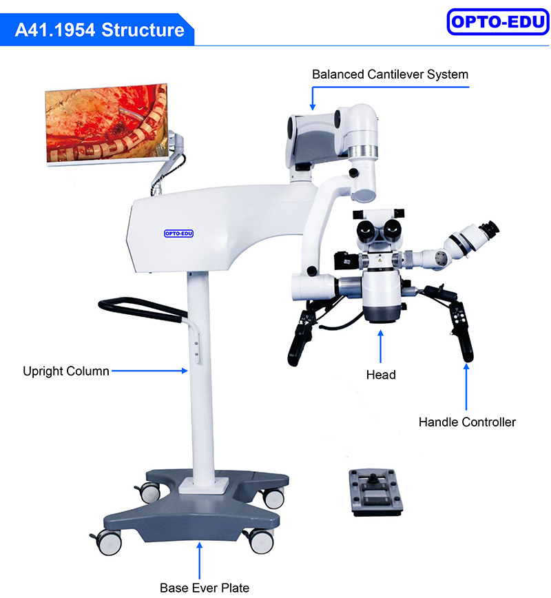
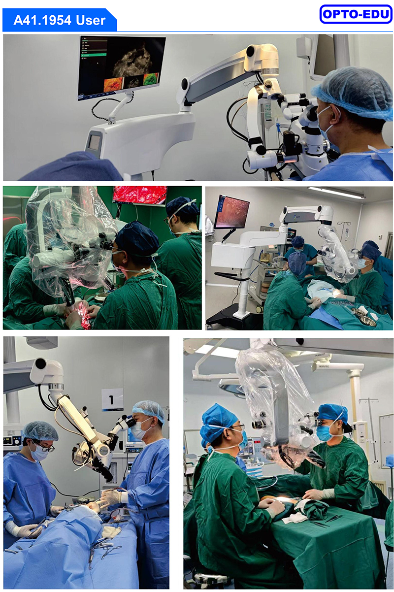
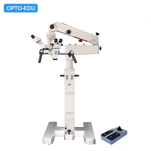
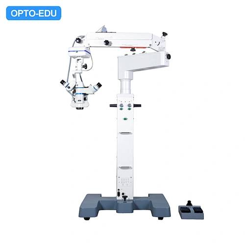
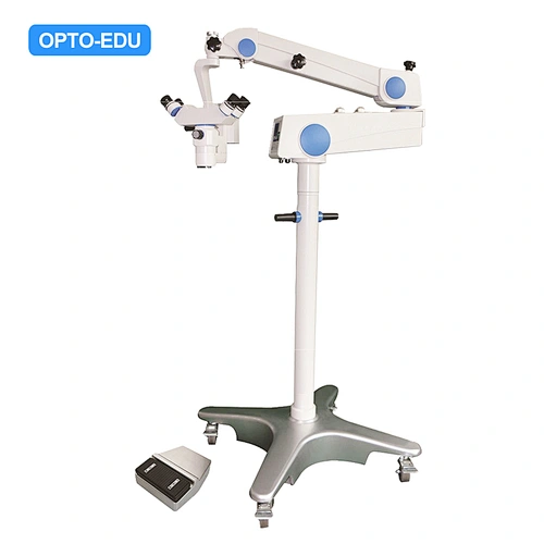
This microscope is mainly used for neurosurgery. ENT、Spine、Ophthalmic、Orthopedics. Surgeons rely on surgical microscopes to visualize the fine anatomical details of the surgical area and brain structure in order to perform the surgical process with high accuracy. It is mainly applied to Brain aneurysm repair, Tumor resections, Arteriovenous malformation (AVM) treatment, Cerebral artery bypass surgery, Epilepsy surgery, Spine surgery. |
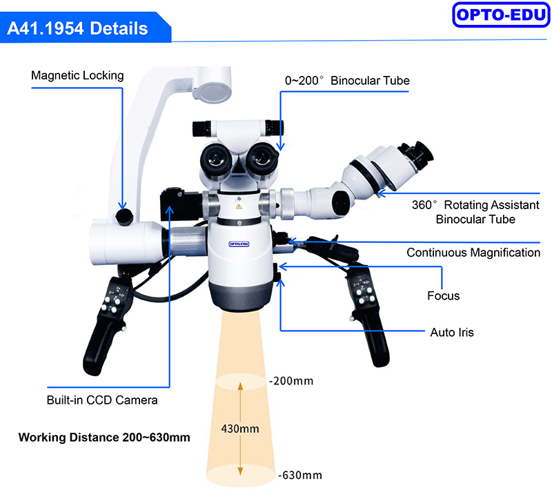
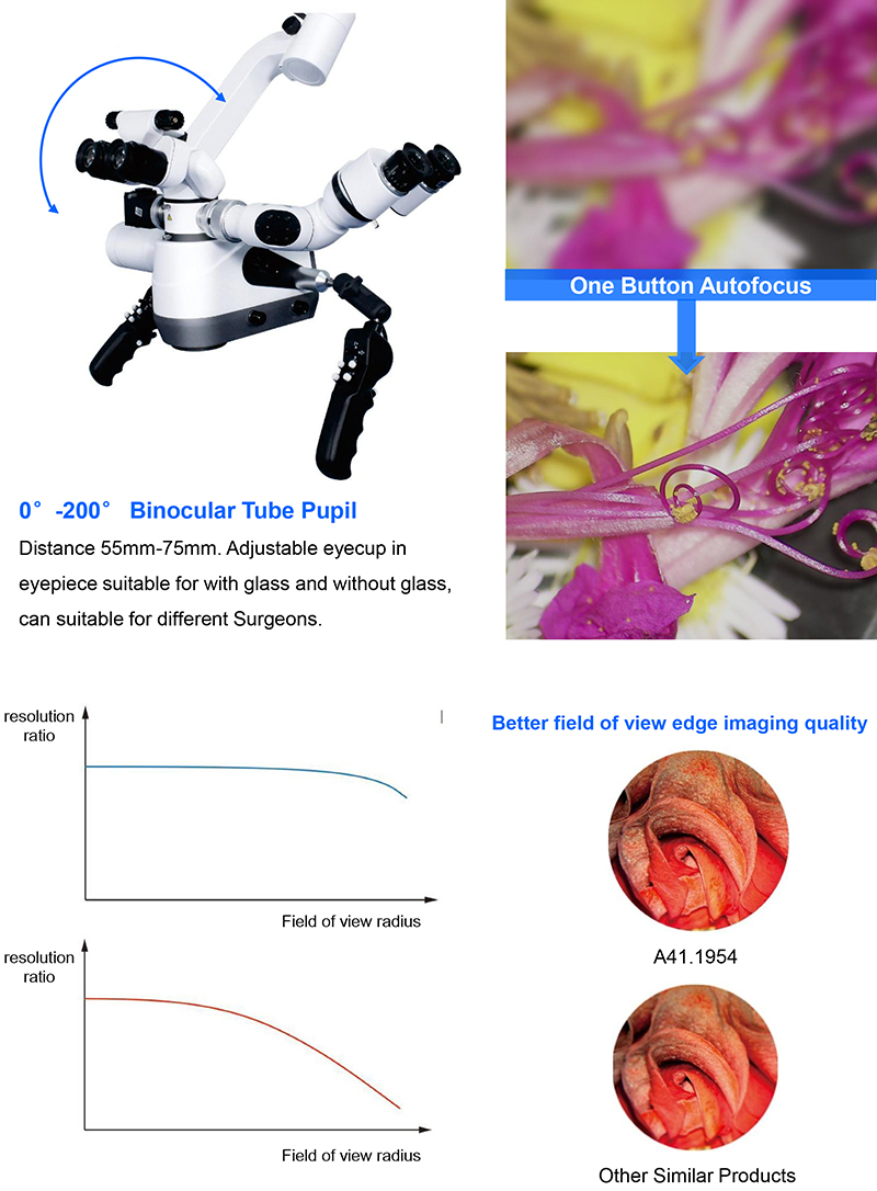
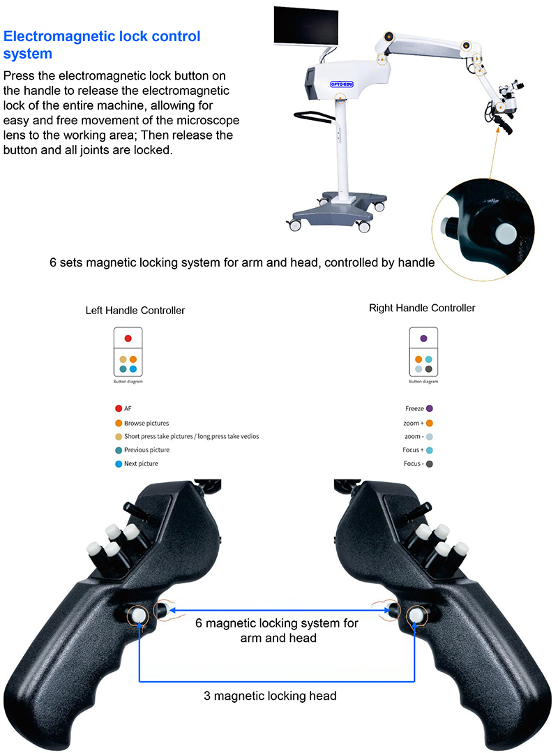
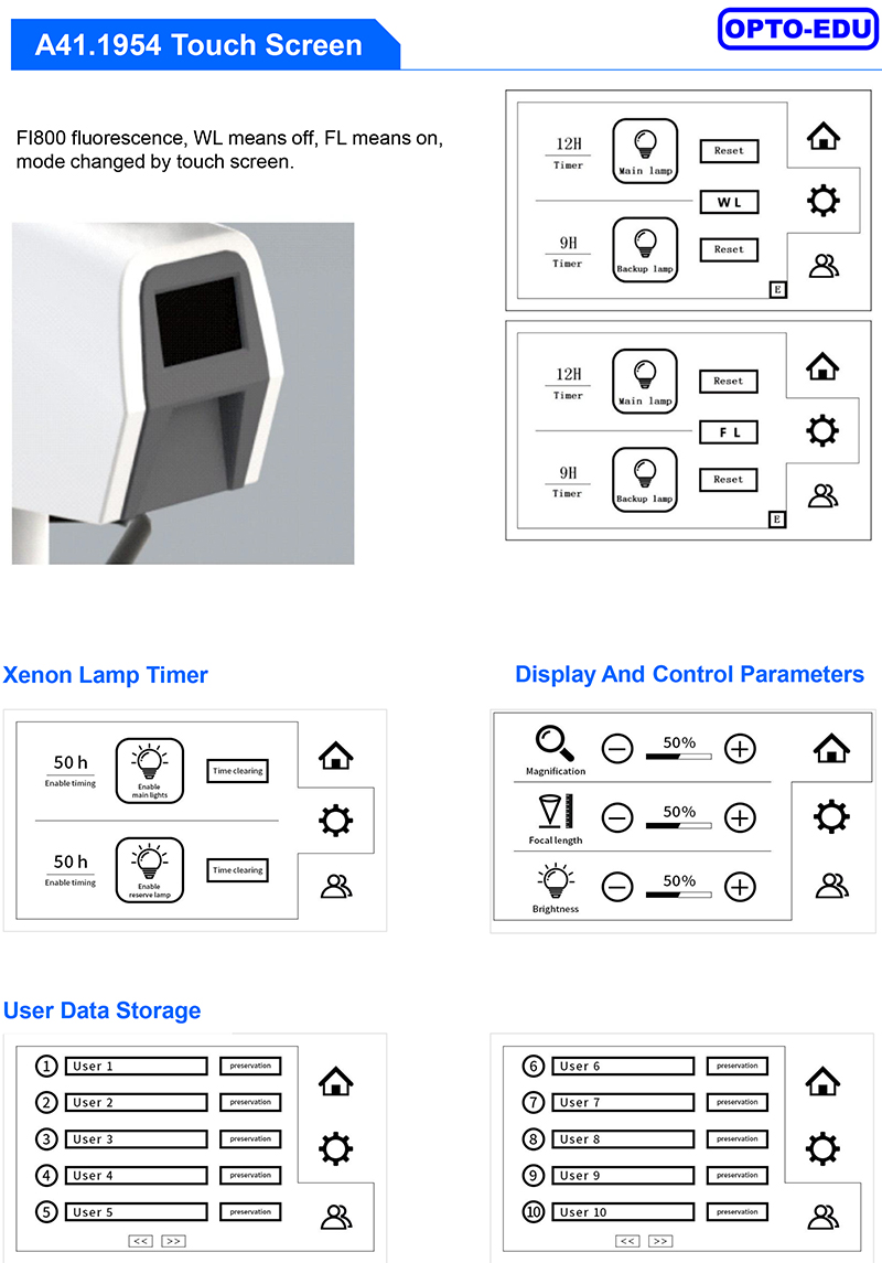
A41.1954 Features -- Best Choice For Neurosurgery, Brain Surgery, Dental, ENT Operation Microscope --6 Sets Magnetic Brake, Eyepiece 12.5x, Motorized Zoom 1.8x~21x, Objective F=200~630mm --Main Binocular Variable Angle 0-200°, With Built-in 4K CCD Camera & Software, Assistant Head 1.8x-21x --12 Function Handle, To Control Zoom, Focusing, Magnetic Lock, Photo/Video, Review, Auto Focus, Brightness Adjust --Built-in Dual Xenon 300W Light, 100000 Lux, Optional FL800, FL560,FL400, X-Y Translator, Face to Face Bridge. |

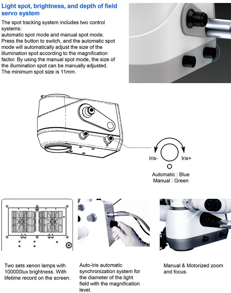
Manual mode: The aperture is controlled by turning the button left and right, and the brightness is controlled by the user. Automatic mode: The aperture decreases as the magnification changes. Automatic mode: The aperture decreases as the magnification increases. Automatic mode: The brightness changes with the focal length: The focal length increases, and the brightness increases. |

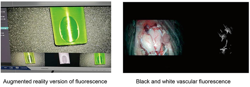
A professional fluorescence camera and fluorescence display software are required. With ICG fluorescence, black and white blood vessel fluorescence can be clearly seen, as well as the flow of indocyanine green reagent in the blood vessels. The enhanced display version of fluorescence (fusion fluorescence) can integrate the fluorescence image with the real-time video image for surgical comparison. |
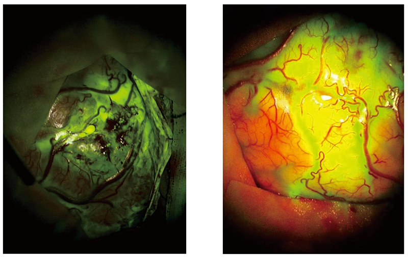
Tumor Fluorescence Equipped with tumor fluorescence imaging, it provides clearer anatomical layers when removing tumors in areas adjacent to important nerves and blood vessels, maximizing the protection of normal functions and reducing surgical complications. |
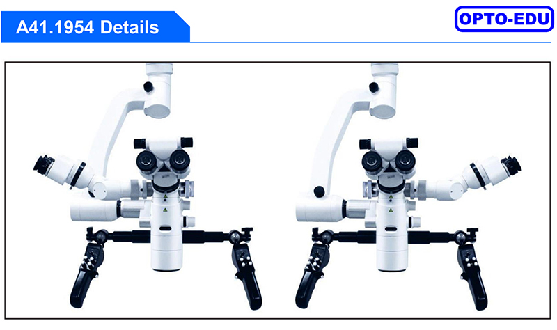
With double-sided splitter, the rotating assistant tube can be on either side , another side directly connect to the camera interface. |
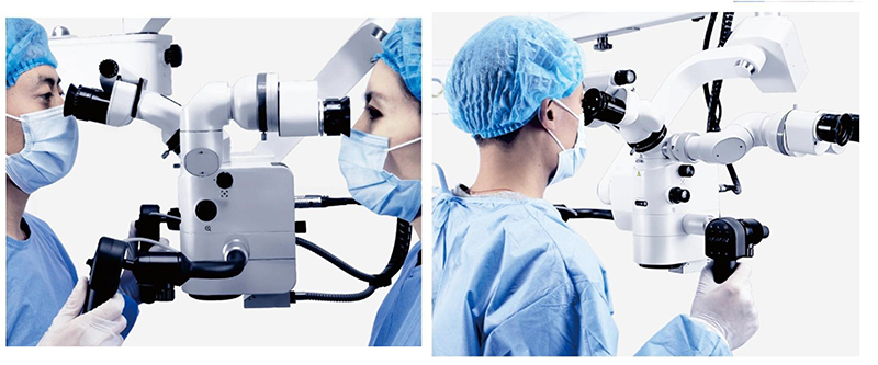
The assistant tube is adjustable, which is 90 degrees and 180 degrees face to face with the Chief surgeon tube, to meet different surgical requirement . |
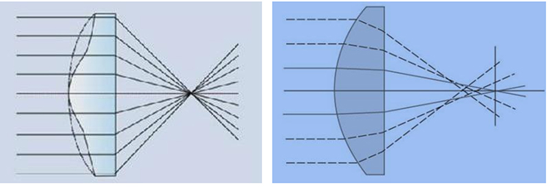
Aspheric Lighting Technology, Better Light Gathering Effect The spherical illumination can not refract the light to the same surface, and cause the light to be more dispersed on the surface. The aspheric technology can increase the concentration effect and improve the brightness. |
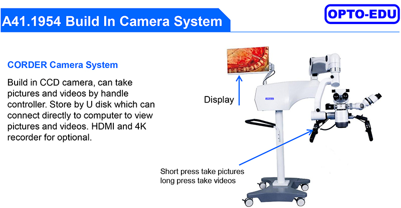
| Image sensor size | 1/1.8inch |
| Image sensor type | CMOS |
| Scanning method | progressive scanning |
| Frame rate | 60fps |
| Effective pixels | 3840(H)x2160(V) |
| Horizontal resolution | ≥1700line |
| Vertical Resolution | ≥1200line |
| Luminance response characteristics | Linear fittingcoefficientR2≥0.99 |
| Signal to Noise Ratio | ≥50dB |
| Spatial frequency response | MTF50:38C/°,MTF30:43C/° |
| Static image tolerance | ≥150 |
| Minimum illumination | 1LUX F1.2 |
| Video Coding | H.265 |
| HDMI | HDMI2.0 |
| DVI | DVI25+4 |
| SDI | 3G |
| LVDS | Dual 8BIT LVDS output |
| Rj45 | 1000M bit/s |
| USB | USB 3.0 |
| Power supply | DC12V 2A |
| Power waste | 7.3W(12V604mA) |
| Operation temperature | 5*C~40*C |
| Working humidity | 30%~85%(No condensing) |
| Atmospheric pressure | 700~1060hPa |
| Output mode | HDMI、DVI、SDI、LVDS RJ45、USB |
| Size | Camera:ϕ25mm:148*100mm |

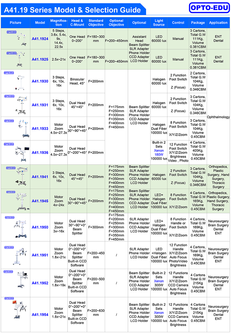
| A41.1950, A41.1951, A41.1952, A41.1954 Surgical Operation Microscope | |||||||
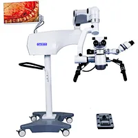
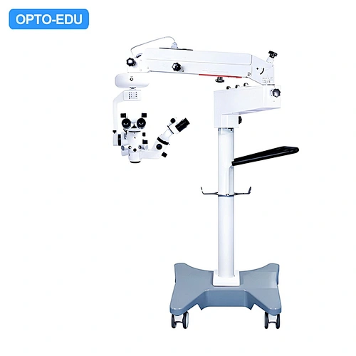
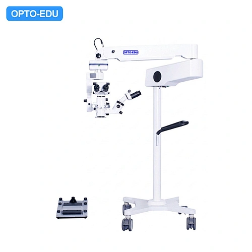
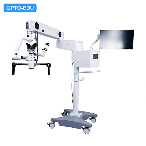
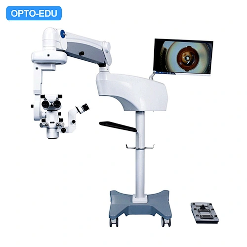
| A41.1950Operating Microscope, is Best Choice for Neurosurgery, Brain Surgery Operation. It has one main head and one assistant head which can rotate 360° freely, Dual Optical fiber coaxial LED + Halogen ,illumination up to 100000 lux, 1:6 Zoom Body, has total magnification 3x~16x, objective F=300mm or 250mm, featured with Motorized Focusing, and 8 Functions foot switch or control handle, it is widely used in hospital for Neurosurgery, Brain Surgery, Spinal, Vascular operation, it can also used for Orthopedics, Hand, Chest, Burn And Plastic Surgery, or Dental, ENT, Andrology, Genitourinary, Animal Anatomy etc. A41.1951is the upgraded version, with built-in CCD camera & software, zoom objective F=200~450mm, 12 functions handle, and dual optical fiber LED + Halogen 100000 Lux light source. A41.1952 is the high-end model in this series, built-in 2 sets Xenon 300W Light, magnetic lock, and 12 functions handle, it can meet most of surgery operation work with best quality & control. A41.1954 is the highest model in this series, built-in 2 sets Xenon 300W Light, magnetic lock, zoom objective F=200~630mm,and 12 functions handle, equipped with Sony 4K 3D monitor , it can meet most of surgery operation work with best quality & control. Features: --Best Choice For Neurosurgery, Brain Surgery, Dental, ENT Operation Microscope --Dual Head Binocular, Eyepiece 12.5x, Motorized Continuous 1:6 Zoom 3x~16x --Main Binocular Variable Angle 30-90°, Assistant's Binocular 30° Can Swing 90° or 270° --8,12 Functions Foot or Handle Control For Zoom, Auto Focus, X-Y Moving, Photo/Video Taking, etc. --Dual Optical Fiber Coaxial Halogen, LED or Xenon Light 100000 lux, Stand Max Extend Range 1452mm | |||||||
| A41.1950 | A41.1951 | A41.1952 | A41.1954 | ||||
| Main Usage | For Neurosurgery, Brain Surgery, Spinal, Vascular | ||||||
| Eyepiece | WF12.5x | ||||||
| Head | Dual Heads For Two People Operation, Assistant Binocular Can Swing 90° to 270° Against Main Head Motorized Swing Head In X Direction +/-45°, In Y Direction +90°, Can Stop At Any Angle Interpupillary Distance 55~75mm, Both Left & Right Eyetube Diopter Adjustable ±6D,A41.1954 Diopter Adjustment ±7D. | ||||||
| Main Head | Inclined 30°~90° | Inclined 0°~200° | |||||
| Magnification | 3x~16x Continuous Zoom | 1.8x~21x Continuous Zoom | 1.8x~19x Continuous Zoom | 1.8x~21x Continuous Zoom | |||
| View Field | Dia.74~12mm | Dia.13~110mm | Dia.12~113mm | Dia. 13.2~180mm | |||
| Zoom Body | 1:6 Motorized Continuous Zoom, Parallel Light System | ||||||
| Assist Head | Inclined 0° | ||||||
| Magnification | 3x~16x Continuous Zoom | 3x~16x Continuous Zoom | 3x~16x Continuous Zoom | 1.8x~21x Continuous Zoom | |||
| View Field | Dia.74~12mm | Dia.74~12mm | Dia.94~13mm | Dia. 13.2~180mm | |||
| Main Obj. | F=300mm | F=200~450mm | F=200~500mm | F=200~630mm | |||
| Assist Obj. | F=180mm | ||||||
| Focus | Motorized Focus, +/-25mm | Motorized Focus 200~450mm | Motorized Focus 200~500mm | Motorized Focus 200~630mm | |||
| Control Switch | 8 Functions Handle or Foot X/Y Moving Z Focusing Zoom | 12 Functions Handle X/Y Moving Z Focusing Zoom Take Photo/Video Review Photo/Video Auto Focus Brightness Adjust | 12 Functions Handle X/Y Moving Z Focusing Zoom CCD Camera Auto Focus Brightness Adjust | 12 Functions Handle X/Y Moving Z Focusing Zoom CCD Camera Auto Focus Brightness Adjust | |||
| Filter | Yellow Filter, Green Filter, Small Spot | ||||||
| Stand | Four Wheels Moving Stand --Max Extended Length 1200mm --Max Rotatable Swing Angle of Arm 0~300° --Height From Objective to Floor 800~1250mm | Four Wheels Moving Stand --Max Extended Length 1715mm --Max Rotatable Swing Angle of Arm 0~300° --Height From Objective to Floor 690~1452mm | |||||
| Lock | Mechanical Lock | Electro Magnetic Lock | |||||
| Illumination | Dual Optical Fiber Coaxial LED + Halogen Light, Light Spot Dia.50mm, Intensity 100000 Lux. | Dual Optical Fiber Coaxial Built-in 2 Sets Xenon300W, Light Spot Dia.50mm, Intensity 100000 Lux. | |||||
| Beam Splitter | Built-in Beam Splitter | ||||||
| Camera, LCD | None | Built-in Camera & Software | Built-in Camera & Software, LCD Monitor | ||||
| Packing | 4 Cartons Total G.W 165Kg Volume 0.45CBM Head:595×460×230(mm)/14KG Body:890×650×265(mm)/41KG Control:1025×260×300(mm)/32KG Stand:890×865×200(mm)/78KG | 4 Cartons Total G.W 215Kg Volume 0.45CBM Head:595×460×230(mm)/14KG Body:890×650×265(mm)/41KG Control:1025×260×300(mm)/32KG Stand:890×865×200(mm)/128KG | |||||




 Huang Xin